Hip artery. Where is the inguinal artery
femoral artery, a. femoralis, is a continuation of the external iliac artery and begins under the inguinal ligament in vascular lacuna. The femoral artery, having entered the anterior surface of the thigh, goes down and medially, lying in the groove between the anterior and medial groups thigh muscles. In the upper third, the artery is located within the femoral triangle, on a deep leaflet of the fascia lata, covered by its superficial leaflet; the femoral vein passes medially from it. Having passed the femoral triangle, the femoral artery (together with the femoral vein) is covered by the sartorius muscle and, at the border of the middle and lower thirds of the thigh, enters the upper opening of the adductor canal. In this canal, the artery is located together with the saphenous nerve, n. saphenus, and femoral vein, v. femoralis. Together with the latter, it deviates posteriorly and exits through the lower opening of the canal to the posterior surface. lower limb into the popliteal fossa, where it receives the name of the popliteal artery, a. poplitea.
The femoral artery gives off a number of branches that supply blood to the thigh and the anterior wall of the abdomen.
1. Superficial epigastric artery, a. epigastrica superficialis, starts from the anterior wall of the femoral artery below the inguinal ligament, pierces the superficial sheet of the broad fascia in the subcutaneous fissure and, rising up and medially, passes to the anterior abdominal wall, where, lying subcutaneously, it reaches the umbilical ring. Here its branches anastomose with the branches of a. epigastrica superior (from a. thoracica interna). Branches of the superficial epigastric artery supply the skin of the anterior abdominal wall and the external oblique muscle of the abdomen. 
2. Superficial circumflex artery ilium, a. circumflexa iliaca superficialis, departs from the outer wall of the femoral artery or from the superficial epigastric artery and goes along the inguinal ligament laterally upward to the superior anterior iliac spine; blood supply to the skin, muscles and inguinal lymph nodes.
3. External genital arteries, aa. pudendae externae, in the form of two, sometimes three thin trunks, are directed medially, bending around the anterior and posterior periphery of the femoral vein. One of these arteries goes up and reaches the suprapubic region, branching out in the skin. Other arteries, passing over the comb muscle, pierce the fascia of the thigh and approach the scrotum (labia) - these are the anterior scrotal (labial) branches, rr. scrotales (labiales) anteriores. 
4. Inguinal branches, rr. inguinales, depart from initial department femoral artery or from the external pudendal arteries (3 - 4) with small trunks and, perforating the wide fascia of the thigh in the area of \u200b\u200bthe ethmoid fascia, supply blood to the skin, as well as superficial and deep lymph nodes of the inguinal region.
5. Deep artery of the thigh, a. profunda femoris, is the most powerful branch of the femoral artery. Departs from its back wall 3 - 4 cm below the inguinal ligament, passes on the iliopsoas and pectineal muscles and goes first outwards, and then down behind the femoral artery. Deviating backwards, the artery penetrates between the vastus medial muscle of the thigh and the adductor muscles, ending in lower third thigh between the large and long adductor muscles in the form of a perforating artery, a. perforans.
The deep artery of the thigh gives off a number of branches.
1) Medial artery, envelope of the femur, a. circumflexa femoris medialis, departs from the deep femoral artery behind the femoral artery, goes transversely inward and, penetrating between the iliopsoas and pectineal muscles into the thickness of the muscles that bring the thigh, goes around the neck from the medial side femur.
The following branches depart from the medial circumflex artery of the femur:
a) ascending branch, r. ascendens, is a small stem, heading up and inward; branching, approaches the comb muscle and the proximal part of the long adductor muscle;
b) transverse branch, r. transversus, - a thin stem, goes down and medially along the surface of the pectinus muscle and, penetrating between it and the long adductor muscle, goes between the long and short adductor muscles; blood supply to the long and short adductor muscles, thin and external obturator muscles;
c) deep branch, r. profundus, is a larger trunk, which is a continuation of a. circumflexa femoris medialis. It goes backwards, passes between the external obturator muscle and the square of the thigh muscle, dividing here into ascending and descending branches;
d) branch of the acetabulum, r. acetabularis, - a thin artery, anastomoses with branches of other arteries supplying the hip joint.
2) The lateral artery enveloping the femur, a, circumflexa femoris lateralis, is a large trunk that departs from the outer wall of the deep artery of the thigh almost at its very beginning. Goes outward in front of the iliopsoas muscle, behind the sartorius muscle and rectus femoris; approaching the greater trochanter of the femur, it is divided into branches: 
a) ascending branch, r. ascendens, goes up and outward, lying under the muscle that stretches the wide fascia and the gluteus medius muscle;
b) descending branch, r. descendens, more powerful than the previous one. Departs from the outer surface of the main trunk and lies under the rectus femoris, then descends along the groove between the intermediate and lateral wide muscles of the thigh. Blood supply to these muscles; reaching the knee area, anastomoses with the branches of the popliteal artery. On its way, it supplies blood to the heads of the quadriceps femoris muscle and gives branches to the skin of the thigh;
c) transverse branch, r. transversus, is a small stem, heading laterally; blood supply to the proximal part of the rectus femoris and the vastus lateralis muscle of the thigh.
3) Perforating arteries, aa. perforantes, usually three, extend from the deep artery of the thigh to various levels and pass to the back of the thigh at the very line of attachment to the femur of the adductor muscles.
The first perforating artery begins at the level of the lower edge of the comb muscle; the second departs at the lower edge of the short adductor muscle and the third - below the long adductor muscle. All three branches pierce the adductor muscles at the place of their attachment to the femur and, having reached the back surface, blood supply the adductor, semimembranosus, semitendinosus muscles, biceps thighs and skin of this area.
The second and third perforating arteries give off small branches to the femur - the arteries feeding the thigh, aa. nutriciae femaris.
4) Descending knee artery, a. descendens genicularis, - a rather long vessel, starts from the femoral artery in the adductor canal, less often - from the lateral artery that envelops the femur. Heading down, perforates along with the saphenous nerve, n. saphenus, from the depth to the surface of the tendon plate, goes behind the sartorius muscle, goes around the inner condyle of the thigh and ends in the muscles of this area and joint capsule knee joint.
This artery gives off the following branches:
a) subcutaneous branch, r. saphenus, into the thickness of the medial wide muscle of the thigh;
b) articular branches, rr. articulares, which take part in the formation of the knee articular network, rete articulare genus, and the patella network, rete patellae.
The femoral artery (a. femoralis) is a continuation of the external iliac artery from the level of the inguinal ligament. Its diameter is 8 mm. In the upper part of the femoral triangle, the femoral artery is located under the lamina cribrosa on the fascia iliopectinea, surrounded by fatty tissue and deep inguinal lymph nodes (Fig. 409). Medial to the artery is the femoral vein. The femoral artery, together with the vein, is medial to m. sartorius in the depression formed by m. iliopsoas and m. pectineus; lateral to the artery lies the femoral nerve. In the middle part of the thigh, this artery is covered by the sartorius muscle. In the lower part of the thigh, the artery, having passed through the canalis adductorius, exits into the popliteal fossa, where it is called the popliteal artery.
Branches of the femoral artery:
1. Superficial epigastric artery (a. epigastrica superficialisis), starting under lig. inguinale, goes to the anterior abdominal wall, supplies it with blood, anastomoses with the superior epigastric artery, which is a branch of a. thoracica interna, with intercostal arteries, with superficial and deep arteries surrounding the ilium.
2. The superficial circumflex iliac artery (a. circumflexa ilium superficialisis) begins with the superficial epigastric artery and reaches the ilium, where it anastomoses with the deep circumflex iliac artery and the branches of the deep femoral artery.
3. External pudendal arteries (aa. pudendae externae), 1-2 in number, depart from medial wall at the level of the beginning of the deep artery of the thigh, pass in the subcutaneous tissue in front of the femoral vein. They supply blood to the scrotum, pubis, in women - large labia.
4. The deep artery of the thigh (a. profunda femoris) has a diameter of 6 mm, departs 3-4 cm below the inguinal ligament from the posterior surface of the femoral artery, forms the medial and lateral branches.
The medial circumflex artery of the femur (a. circumflexa femoris medialis) starts from the posterior wall of the deep artery of the thigh and after 1–2 cm is divided into superficial, deep transverse and acetabular branches. These branches supply blood to the adductor muscles of the thigh, the obturator and square muscles, the neck of the femur, and the articular bag. The artery anastomoses with the obturator, inferior gluteal, and lateral arteries surrounding the femur.
The lateral circumflex artery of the femur (a. circumflexa femoris lateralis) originates from the lateral wall of the deep artery of the thigh and after 1.5-3 cm is divided under m. sartorius and m. rectus femoris into ascending, descending and transverse branches. The descending branch is more developed than the others and supplies blood to the anterior muscles of the thigh. The ascending branch, passing under m. rectus femoris and m. tensor fasciae latae), wraps around the femoral neck and anastomoses with the medial artery. The transverse branch supplies blood to the muscles of the middle part of the thigh.
Perforating arteries (aa. perforantes), 3-4 in number, are the terminal branches of the deep artery of the thigh. They pass to the back of the thigh through m. adductor longus et magnus. They supply blood to the adductor and posterior muscles of the thigh, the femur. Anastomose with the branches of the deep femoral artery, superior and inferior gluteal and obturator arteries listed above.
5. The descending knee artery (a. genus descendens) starts from the terminal part of the femoral artery within the adductor canal of the thigh (canalis adductorius). Together with n. saphenus leaves the canal above the knee joint from the medial side. It supplies blood to the medial head of the quadriceps femoris muscle, the joint capsule. Anastomoses with branches of the popliteal artery.
The femoral artery is the largest vessel that supplies blood to:
- muscles and skin of the anterior abdominal wall;
- nodes of the inguinal region and tissue of the Scarpa triangle;
- thigh muscles;
- hip bones;
- reproductive system;
- calf and ankle muscles.
Capillaries are intermediaries. Delivering oxygen and nutrients to all areas of the body. The diameter of the artery is about 8 mm. The femoral continues the iliac, from the level of the inguinal ligament, where it branches.
The combination of the epigastric, superficial femoral and external pudendal arteries form the triangle of Scarpa. FROM inside this area is surrounded by muscles and inguinal ligaments, outside - thin skin, where the pulsation is clearly palpable. Here the artery is clamped during femoral bleeding.
The location of the artery is the tendon canal in the thigh with an exit in the popliteal fossa, where a clear pulsation is also felt. According to its structure and location, the femoral artery and the accompanying vascular system in each person may have minor differences that do not affect general functions blood supply.
Atherosclerosis of the femoral artery
Atherosclerosis is a chronic lesion of an artery that occurs as a result of the appearance of cholesterol deposits that pollute the inner walls of blood vessels. Consequence: the lumen in the vessels gradually narrows and occurs oxygen starvation organs, impaired peripheral circulation. Untimely treatment can lead to complete blockage of blood vessels or rupture of the artery. Also, malnutrition can lead to necrosis (gangrene).
A fatal outcome is observed with untimely treatment in 30% within 5 years from the onset of the pathology.
Causes of pathology
As a rule, atherosclerosis of the femoral artery occurs more often in males, the elderly (after 65 years). Also at risk of the disease are persons whose relatives have hyperlipidemia (high blood fat).
- with high blood pressure;
- diabetes mellitus;
- hyperlipidemia;
- availability bad habits(smoking, excessive alcohol consumption);
- injuries;
- depression.
A sedentary lifestyle and overweight are a direct path to atherosclerosis of the femoral artery and not only ...
Symptoms
Vivid symptoms in atherosclerosis are observed only in 10 patients out of a hundred. In some cases, there are no signs of pathology.
- pain in the legs when walking or increased physical activity(may cause lameness). Syndromes disappear during a break in activity or rest;
- numbness, weakness, tingling in the legs when walking;
- aching pain and burning sensation in the legs during the rest period after physical exertion;
- ulcers, corns, which are accompanied by pain in the legs and feet;
- coldness in the legs;
- change in skin color (with critical ischemia);
- hair loss in the shin area;
- loss of muscle strength and energy.
Diagnostics
Initially, the specialist performs an external examination, during which the following are observed:
- thickening and shine skin;
- alopecia on the affected areas;
- fragility of nails;
- change in skin color;
- thinning of the muscles of the diseased limb.
With the help of palpation, skin temperature, pulsation are determined, sensitivity and motor activity are also determined.
With the help of modern equipment, the diagnosis is clarified and the most effective treatment. Specialists resort to:
- dopplerography or duplex scanning. The method has high accuracy and is based on the use of ultrasound capabilities;
- CT angiography, which is a type of X-ray examination, during which the patient is irradiated;
- MR angiography using magnetic resonance imaging. In this case, the image of a blood vessel is studied;
- standard angiography - the usual fluoroscopic examination of the artery using radiopaque agents.
Diagnostics carried out by professional methods will be the key to successful treatment of atherosclerosis
A puncture of the femoral artery is performed to obtain a blood sample, direct measurement of blood pressure, injection of a contrast agent with certain research methods.
Treatment
The treatment of atherosclerosis combines drug therapy, exercise, a healthy diet, and getting rid of disease-causing factors. Application folk remedies can also be included in therapy, but as additional method.
Physical activity is provided by special training 3 times in 7 days for an hour. Training walking has a good effect.
Vascular complications are reduced with the help of antiplatelet therapy (drugs Aspirin and Clopidogrel).
The permeability of blood through the femoral artery increases with the use of phosphodiesterase inhibitors (Pletala and others).
The operation is prescribed for advanced disease, its progression or ineffective conservative treatment.
The type of surgical treatment is prescribed by the doctor depending on clinical picture pathology. Experts resort to the following methods:
- Balloon angioplasty. The method consists in introducing a catheter with a miniature balloon through a puncture in the skin. Then the balloon is inflated and the atherosclerotic plaque is “crushed”. To achieve the best effect, balloon angioplasty and stenting are used together.
- Prosthetics. A piece of vein or prosthesis replaces the blocked vessel.
- Shunting. During surgery, an additional path for blood is created, which bypasses the affected area.
- Endarterectomy. Represents an open surgical operation, during which not only the cholesterol plaque is removed, but also the affected lining of the artery wall.
- Stenting. A stent (a metal mesh tube) is inserted into the narrowed artery, which prevents the vessel from narrowing.
Thrombosis
Thrombosis of the femoral artery is formed by blood clots that provoke stenosis and blockage of the vessel. This disease differs from atherosclerosis, in which cholesterol formations are observed. Often, atherosclerosis is the cause of thrombosis.
The following factors lead to thrombosis:
- vascular damage (postponed chemotherapy, improperly installed venous catheter or unprofessional injection into a vein, injury, etc.);
- reduced speed of blood movement through the vessels (pregnancy, overweight, varicose veins, etc.);
- increased blood clotting (delivery, pregnancy, dehydration, surgery, diabetes);
- high levels of cholesterol in the body.
Thrombosis after sixty is a common thing
Symptoms
With thrombosis, the patient complains:
- on pulling or arching pain in the calf muscles and feet. The development of the disease contributes to the intensification and frequency of attacks of pain. There is an inability of the patient to walk for a long time, he constantly needs rest;
- swelling and numbness of the legs;
- increase in body temperature;
- pallor of the skin of the affected area.
The diagnosis of thrombosis is identical to the diagnosis of atherosclerosis.
Treatment
If the thrombus is in a stable state, the risk of separation is minimal, or there are contraindications to surgical intervention, specialists resort to drug treatment:
- antithrombotic therapy, the purpose of which is to destroy and prevent the growth of a blood clot;
- anticoagulation therapy, which thins the blood and normalizes its composition;
- restoration of effective blood circulation.
To improve the outflow of blood, the patient is bandaged limbs elastic bandage.
Aneurysm
Aneurysm of the femoral artery is the most common pathology. It is expressed in a saccular protrusion of the artery wall, observed in a small area or, conversely, affecting a large area. This anomaly is formed as a result of loss of elasticity and thinning of the vessel wall due to:
- the presence of atherosclerotic plaques;
- hypertension;
- infectious diseases (vasculitis);
- previous operations.
Sports injuries often cause aneurysms
Risk factors include the presence of infections in the body, overweight, and heredity.
Symptoms of an aneurysm are similar to those of thrombosis. The difference lies in the presence of an elastic pulsating seal on the affected area.
Aneurysm is not treatable with medicines and methods traditional medicine. At the initial stages, specialists monitor the development of the disease, in severe cases they resort to shunting, vessel replacement or stenting.
False aneurysm
False aneurysm is observed in case of tissue injuries that cause damage to the vessel. The accumulation of blood in damage to the walls of the vessel creates a pulsating hematoma.
Vascular wall damaged:
- in case of a poorly performed medical injection in the course of therapeutic or diagnostic measures;
- purulent inflammatory processes in tissues in close proximity to the vessel, leading to violation of the walls of the vessel, bleeding and the formation of a hematoma;
- injuries.
A false aneurysm leads to the following symptoms:
- growing swelling in the affected area;
- pain sensations of a different nature;
- change in skin color;
- pulsations.
If the false aneurysm is small, it usually goes away on its own.
In other cases, specialists resort to endovascular, compression methods or surgical intervention.
Embolism
Embolism of the femoral artery - the presence in the arterial bed of emboli (pieces of a blood clot, fatty accumulations and other foreign bodies) that move through the vessel and cause occlusion.
Emboli in the arteries of the lower extremities
The overall picture is expressed in a pronounced character:
- acute pain;
- blanching of the skin with the subsequent appearance of cyanosis;
- marbling of the skin;
- lowering the temperature of the affected limbs;
- sensitivity disorder.
In the diagnosis of pathology, the absence of pulsation at the site of the lesion is revealed. The most informative in this case is the method of angiography.
Best effect wears surgery, then heparin therapy and getting rid of the diseases that caused the pathology.
Each of the above pathologies can lead to irreversible consequences. To avoid this, you need to follow simple rules: lead an active lifestyle, eat right, undergo regular medical examination and avoid injury.
Anatomy of the femoral artery
The femoral artery (FA) in anatomy is a blood vessel originating from the external iliac trunk. The connection of these two channels occurs in the human pelvis. The diameter of the barrel is 8 mm. What branches does the common femoral artery consist of and where are they located?
Location
The femoral artery originates from the iliac trunk. On the outer side of the leg, the channel extends down into the groove between the muscle tissues.
A third of its upper part is located in the triangle of the thigh, where it is located between the sheets of the femoral fascia. A vein runs next to an artery. These vessels are protected by sartorial muscle tissue, they go beyond the borders of the femoral triangle and enter the opening of the adductor canal located from above.
In the same place there is a nerve located under the skin. The femoral branches go back a little, moving through the canal opening, go to the back of the leg and enter the area under the knee. At this site, the femoral canal ends and the popliteal artery begins.
main branches
Several branches depart from the main blood trunk, which supply blood to the femoral part of the legs and the anterior surface of the peritoneum. Which branches are included here can be seen in the following table:
In this place, it stretches under the skin, reaching the navel, it merges with other branches. The activity of the epigastric superficial artery is to provide blood to the skin, the walls of the external oblique muscle tissues of the abdomen.
The remaining branches move over the comb muscle, pass through the fascia and go to the genitals.
inguinal branches
They originate from the external genital arteries, after which they reach the wide femoral fascia. PV provide blood supply to the skin, tissues, lymph nodes located in the groin.
deep femoral artery
It starts at the back of the joint, just below the groin. This branch is the largest. The vessel stretches through the muscle tissues, first goes outward, then goes down behind the femoral artery. Then the branch moves between the muscles of the area under consideration. The trunk ends approximately in the lower third of the thigh, goes to the perforating arterial canal.
The vessel enveloping the femur leaves the deep trunk, heading into the depths of the limb. After that, it passes near the neck of the femoral bone.
Branches of the medial canal
The medial artery has its own branches that run around the femur. Branches include:
- Rising. It is presented in the form of a small trunk that runs in the upper and inner parts. Then several more branches depart from the vessel, heading towards the tissues.
- Transverse. Thin, goes to the lower zone along the surface of the comb muscle to pass between it and the adductor muscle tissue. The vessel provides blood to nearby muscles.
- Deep. It is the largest in size. Moves to the back of the thigh, passes between the muscles and branches into two components.
- Vessel of the acetabulum. This is a thin branch that enters other arteries of the lower extremities. Together they supply blood to the hip joint.
Lateral trunk
The lateral artery goes around the femoral bone, leaves the surface of the deep canal outward.
After that, it is removed to the outer region of the anterior iliopsoas, posterior sartorius and rectus muscles. Approaches the greater trochanter of the thigh bone and breaks up into:
- Ascending branch. Moves to the top, goes under the tissue surrounding the fascia of the thigh, and the gluteal muscle.
- descending branch. Is powerful enough. It starts from the outer wall of the main trunk, runs under the rectus femoral muscle, goes down between the tissues of the legs, nourishing them. Then it reaches the knee zone, connects with the branches of the artery located under the knee. Passing through the muscles, it supplies blood to the quadriceps femoral muscle, after which it is divided into several branches moving towards the skin of the limb.
- Cross branch. It is presented in the form of a small trunk. The vessel supplies the proximal part of the rectus and lateral muscle tissue.
Perforating channels
There are only 3 such trunks. They start from the deep femoral artery in its different parts. Vessels move to the back wall of the thigh at the place where the muscles connect to the bone.
The first perforating vessel departs from the lower zone of the pectineus muscle, the second from the short, and the third from the long adductor tissue. These vessels pass through the muscles at the junction with the thigh bone.
Then the perforating arteries go towards the posterior femoral surface. They provide blood to the muscles and skin in this part of the limb. There are several other branches from them.
Descending artery of the knee
This vessel is very long. It starts from the femoral artery in the adductor canal. But it can also depart from the lateral vessel, which goes around the thigh bone. This is much less common.
The artery descends, intertwines with the nerve under the skin, then goes to the surface of the tendon plate, passes from the back of the tailor's fabric. After that, the vessel moves around the internal femoral condyle. It ends in the muscles and the knee joint.
The descending trunk of the knee has the following branches:
- Subcutaneous. It is located deep in the medial wide tissue of the limb.
- Articular. This femoral branch is involved in the formation of a network of joints of the knee and patella.
Vascular disorders
There are a large number of different pathologies that affect the circulatory system, which leads to disruption of the body. The branches of the artery of the femoral part are also exposed to diseases. The most common of them are:
- Atherosclerosis. This disease is characterized by the formation of cholesterol plaques in the vessels. The presence of this pathology increases the risk of thromboembolism. A large accumulation of deposits causes weakening and damage to its wall, impairs patency.
- Thrombosis. The disease is the formation of blood clots that can lead to dangerous consequences. If a blood clot blocks the vessel, then the tissues of the legs will begin to die. This leads to limb amputation or death.
- Aneurysm. The disease is no less dangerous for the life of patients. With it, a protrusion occurs on the surface of the artery, the vessel wall becomes thinner and more vulnerable to damage. A ruptured aneurysm can be fatal due to rapid and massive blood loss.
These pathological conditions proceed without clinical manifestations in the early stages, making it difficult to detect them in a timely manner. Therefore, it is necessary to check regularly for circulatory problems.
If one of the pathologies is detected, the treatment regimen should be prescribed exclusively by a doctor. Under no circumstances should these violations be ignored.
Thus, the femoral artery has a complex structure, a large number of branches. Each vessel performs its role, supplying the skin and other parts of the lower limb with blood.
Femoral artery: structure, functions, anatomy
Anatomy is a science that studies the human structure. In this article, we will consider the femoral artery, its location and main branches.
Location
The femoral artery departs and continues the external iliac artery, originates in the vascular lacuna under the inguinal ligament. On the outer surface of the thigh, it moves down and is medially located in the groove between the muscle groups (anterior and medial). Its upper third is located in the femoral triangle, located on a sheet of a wide fascia, covered from above by its surface sheet; on the medial side, it is adjacent to the femoral vein.
Having gone beyond the femoral triangle, the femoral artery and vein, which are covered by the sartorius muscle, approximately at the border of the lower and middle thirds of the thigh, enter the afferent canal, its upper opening. Here, in the canal, is the saphenous nerve and, as already mentioned, the femoral vein. The artery and vein deviate posteriorly, pass through the lower canal opening, following to the lower limb (its posterior surface), descending into the popliteal fossa, where they pass into the popliteal artery.
Where is the femoral artery located in humans? This question is often asked. Let's consider it in more detail in this article.
Main branches of the femoral artery
Several branches that provide blood supply to the thigh and abdominal wall in front depart from the femoral artery. What are these branches?
The epigastric superficial artery branches off from the femoral artery, or rather, its anterior wall, in the region of the inguinal ligament, deepens into the superficial sheet of the fascia lata, then rises up and medially, passing to the anterior abdominal wall. Passing subcutaneously, it reaches the umbilical ring, where it anastomizes (merges) with several more branches. The main function of the branches of the superficial epigastric artery is the blood supply to the skin of the abdominal wall in front and the external oblique muscles of the abdomen.
The superficial femoral artery, bending around the ilium, moving away from the superficial epigastric artery, rushes laterally and upwards parallel to the inguinal fold reaches the anterior superior iliac bone; provides blood supply to the skin, muscles and inguinal lymph nodes.
The external genital arteries, most often there are two or three stems, have a medial direction, go around the periphery of the femoral vein (posterior and anterior). Then one of the arteries, heading up, reaches the area above the pubis and branches in the skin. The other two pass over the comb muscle, perforating the fascia of the thigh, rush to the labia (scrotum). These are the so-called anterior labial (scrotal) branches.
They make up the femoral artery. Her anatomy is unique.
inguinal branches
The inguinal branches in small trunks depart from the external genital arteries (the initial section of the femoral artery), then pass the wide fascia of the thigh in the region of the ethmoid fascia, supply blood to the deep and superficial lymphatic inguinal nodes, as well as the skin.
deep femoral artery
The deep femoral artery, starting from its posterior wall, approximately 3-4 cm lower than the inguinal ligament, passes through the comb and iliopsoas muscles, goes outward at the beginning, and then down, located behind the femoral artery. This is her biggest thread. After the artery follows between the adductor muscles and the wide medial muscle of the thigh, and its end is approximately the lower third of the thigh between the long and large adductor muscles with the transition to the perforating artery.
These are the numerous branches of the femoral artery.
Bending around the femur, the medial artery, moving away from the deep and behind the femoral artery, goes inward, penetrating transversely into the thickness of the crest and iliopsoas muscles adducting the thigh, then goes around the neck of the femur from the medial side.
Branches from the medial artery
The following branches depart from the medial artery:
- the ascending branch is a small stem that has an upward and inward direction; branching when approaching the pectinate and long adductor (proximal) muscles;
- the transverse branch passes medially and down the surface of the pectinus muscle, passing between the long adductor and pectineus muscle, then between the long and short adductor muscles; provides blood supply to the long and short adductor muscles, thin and external obturator muscles.
- deep branch - a relatively large trunk, is a continuation of the medial artery. It has a posterior direction, passing between the square and external obturator muscle, then it is divided into descending and ascending branches;
- a branch of the acetabulum, a small artery that anastomoses with branches of other arteries, supplies blood to the hip joint. It is here that the pulsation of the femoral artery is felt.
Lateral artery
The lateral circumflex femoral artery is a very large vessel that branches off almost at the very beginning of the deep femoral artery, from its outer wall. Directed outward, passes in front of the iliopsoas muscle, but behind the rectus and sartorius muscles of the thigh, and is divided when the greater trochanter of the femur is reached.
a) the ascending branch passes under the muscle that stretches the fascia lata and the gluteus medius; has an upward and outward direction.
b) the descending branch is more powerful than the previous branch. Departs from the outer surface of the main trunk, passes under the rectus femoris muscle, descends along the groove located between the lateral and intermediate wide muscles of the thigh. Supplies these muscles with blood. Anastomoses in the knee region with branches of the popliteal artery. Along the way, it supplies blood to the head of the quadriceps femoris muscle, and also branches off to the skin.
c) transverse branch - a small trunk that supplies blood to the rectus muscle (its proximal part) and the lateral wide muscle of the thigh, the direction is lateral.
perforating arteries
Three perforating arteries depart at different levels from the deep femoral artery, then pass to the posterior surface of the thigh, in the region of attachment of the adductor muscles to the femur. The beginning of the first perforating artery is at the level of the lower edge of the pectinate muscle; the second begins at the short adductor muscle (lower edge), and the third below the adductor muscle long. Having passed through the adductor muscles, at the places where they are attached to the femur, all three branches find an exit at the back surface. Produce blood supply to the following muscles: adductor, semimembranosus, semitendinosus, biceps femoris, and skin in this area.
From the second and third branches, in turn, depart small branches that feed the femur of the perforating artery.
Descending genicular artery
The descending genicular artery is a very long vessel that originates from the femoral artery inside the adductor canal (sometimes it originates from the lateral artery that goes around the femur). It descends along with the saphenous nerve, under the tendon plate, passes behind the sartorius muscle, then bypasses the internal condyle of the thigh and ends in the thickness of the muscles of this area and the capsule of the knee joint.
The following branches are given by the above artery:
- subcutaneous branch, supplying the medial part of the wide muscle of the thigh;
- articular branches that form the knee articular network of vessels, and networks of the patella.
We examined the femoral artery, its anatomical structure.
Anatomy and function of the superficial femoral artery
The superficial femoral artery is one of the branches of a large vessel of the lower extremities, extending from the external iliac artery.
Let us consider in more detail the anatomy of the femoral artery, which is conditionally divided into two parts:
- General - passing from the inguinal ligament to the area of \u200b\u200bbifurcation (division). One of the large branches of the common femoral artery is the superficial epigastric artery, which gives off small vessels that feed the external genitalia and thigh structures. It passes through the cribriform fascia into subcutaneous tissue and goes to the anterior wall of the peritoneum, anastomosing with the internal thoracic artery.
- Superficial - starting in the bifurcation zone of the common femoral artery.
The last branch, bending around the ilium, runs laterally towards the superior anterior iliac spine, being parallel to the inguinal fold. In adjacent muscle structures, skin, and lymph nodes, the superficial femoral artery is connected by an orifice to the deep femoral artery, which is the largest branch.
It departs from the posterior semicircle of the femoral artery, just below the inguinal ligament (3-4 cm), dividing into the medial, lateral and perforating arteries. Functions: is the main source of blood supply to the thigh.
The superficial femoral artery branches into a number of small vessels. A large descending artery of the knee also departs from it, which takes the main part in the formation of the vascular arterial network of this element of the lower limb. This branch separates in the adductor canal, heading through the tendon gap of the adductor muscle to the front of the thigh along with the saphenous nerve.
The superficial femoral artery, deviating posteriorly in the lower third, enters the femoropopliteal canal, which is the adductor muscles and ligaments of the thigh. Then the vessel exits the canal and continues into the popliteal artery. The latter, located in the popliteal fossa, gives several small branches that connect with each other and form the knee arterial network. In the area where the anterior tibial artery departs, the popliteal artery ends, anastomosing into the posterior tibial artery.
Examination of the vessels of the thigh
To study the characteristics of the femoral artery and all its branches, as well as to assess their condition and identify possible pathological abnormalities, it is recommended to use a linear probe with a frequency of 5 MHz. It is important that the superficial femoral artery can be traced quite well almost throughout, namely to the lower third of the thigh - the area of its entry into the femoral-popliteal canal. To conduct a study of this vessel, the patient should be in the supine position, straightening and slightly moving the legs.
Arteries of the lower limb. Femoral artery.
Femoral artery, a. femoralis, is a continuation of the external iliac artery and begins under the inguinal ligament in the vascular lacuna. The femoral artery, having entered the anterior surface of the thigh, goes down and medially, lying in the groove between the anterior and medial thigh muscle groups. In the upper third, the artery is located within the femoral triangle, on a deep leaflet of the fascia lata, covered by its superficial leaflet; the femoral vein passes medially from it. Having passed the femoral triangle, the femoral artery (together with the femoral vein) is covered by the sartorius muscle and, at the border of the middle and lower thirds of the thigh, enters the upper opening of the adductor canal. In this canal, the artery is located together with the saphenous nerve, n. saphenus, and femoral vein, v. femoralis. Together with the latter, it deviates backwards and exits through the lower opening of the canal to the posterior surface of the lower limb into the popliteal fossa, where it receives the name of the popliteal artery, a. poplitea.
1. Superficial epigastric artery, a. epigastrica superficialis, starts from the anterior wall of the femoral artery below the inguinal ligament, pierces the superficial sheet of the broad fascia in the subcutaneous fissure and, rising up and medially, passes to the anterior abdominal wall, where, lying subcutaneously, it reaches the umbilical ring. Here its branches anastomose with the branches of a. epigastrica superior (from a. thoracica interna). Branches of the superficial epigastric artery supply the skin of the anterior abdominal wall and the external oblique muscle of the abdomen.
2. Superficial artery circumflexing the ilium, a. circumflexa iliaca superficialis, departs from the outer wall of the femoral artery or from the superficial epigastric artery and goes along the inguinal ligament laterally upward to the superior anterior iliac spine; blood supply to the skin, muscles and inguinal lymph nodes.
3. External genital arteries, aa. pudendae externae, in the form of two, sometimes three thin trunks, are directed medially, bending around the anterior and posterior periphery of the femoral vein. One of these arteries goes up and reaches the suprapubic region, branching out in the skin. Other arteries, passing over the comb muscle, pierce the fascia of the thigh and approach the scrotum (labia) - these are the anterior scrotal (labial) branches, rr. scrotales (labiales) anteriores.
4. Inguinal branches, rr. inguinales, depart from the initial section of the femoral artery or from the external pudendal arteries (3-4) with small trunks and, perforating the wide fascia of the thigh in the area of the ethmoid fascia, supply the skin, as well as the superficial and deep lymph nodes of the inguinal region.
5. Deep artery of the thigh, a. profunda femoris, is the most powerful branch of the femoral artery. Departs from its back wall 3 - 4 cm below the inguinal ligament, passes on the iliopsoas and pectineal muscles and goes first outwards, and then down behind the femoral artery. Deviating backwards, the artery penetrates between the vastus medial muscle of the thigh and the adductor muscles, ending in the lower third of the thigh between the large and long adductor muscles in the form of a perforating artery, a. perforans.
The deep artery of the thigh gives off a number of branches.
1) Medial artery, envelope of the femur, a. circumflexa femoris medialis, departs from the deep femoral artery behind the femoral artery, goes transversely inward and, penetrating between the iliopsoas and pectineal muscles into the thickness of the muscles that bring the thigh, goes around the femoral neck from the medial side.
a) ascending branch, r. ascendens, is a small stem, heading up and inward; branching, approaches the comb muscle and the proximal part of the long adductor muscle;
b) transverse branch, r. transversus, - a thin stem, goes down and medially along the surface of the pectinus muscle and, penetrating between it and the long adductor muscle, goes between the long and short adductor muscles; blood supply to the long and short adductor muscles, thin and external obturator muscles;
c) deep branch, r. profundus, is a larger trunk, which is a continuation of a. circumflexa femoris medialis. It goes backwards, passes between the external obturator muscle and the square of the thigh muscle, dividing here into ascending and descending branches;
d) branch of the acetabulum, r. acetabularis, - a thin artery, anastomoses with branches of other arteries supplying the hip joint.
2) The lateral artery enveloping the femur, a, circumflexa femoris lateralis, is a large trunk that departs from the outer wall of the deep artery of the thigh almost at its very beginning. Goes outward in front of the iliopsoas muscle, behind the sartorius muscle and rectus femoris; approaching the greater trochanter of the femur, it is divided into branches:
a) ascending branch, r. ascendens, goes up and outward, lying under the muscle that stretches the wide fascia and the gluteus medius muscle;
b) descending branch, r. descendens, more powerful than the previous one. Departs from the outer surface of the main trunk and lies under the rectus femoris, then descends along the groove between the intermediate and lateral wide muscles of the thigh. Blood supply to these muscles; reaching the knee area, anastomoses with the branches of the popliteal artery. On its way, it supplies blood to the heads of the quadriceps femoris muscle and gives branches to the skin of the thigh;
c) transverse branch, r. transversus, is a small stem, heading laterally; blood supply to the proximal part of the rectus femoris and the vastus lateralis muscle of the thigh.
3) Perforating arteries, aa. perforantes, usually three, depart from the deep artery of the thigh at various levels and pass to the back of the thigh at the very line of attachment to the femur of the adductor muscles.
The first perforating artery begins at the level of the lower edge of the comb muscle; the second departs at the lower edge of the short adductor muscle and the third - below the long adductor muscle. All three branches pierce the adductor muscles at the place of their attachment to the femur and, having reached the posterior surface, blood supply the adductor, semimembranosus, semitendinosus, biceps femoris and skin of this area.
The second and third perforating arteries give off small branches to the femur - the arteries feeding the thigh, aa. nutriciae femaris.
4) Descending knee artery, a. descendens genicularis, - a rather long vessel, starts from the femoral artery in the adductor canal, less often - from the lateral artery that envelops the femur. Heading down, perforates along with the saphenous nerve, n. saphenus, from the depth to the surface of the tendon plate, goes behind the sartorius muscle, goes around the inner condyle of the thigh and ends in the muscles of this area and the articular capsule of the knee joint.
a) subcutaneous branch, r. saphenus, into the thickness of the medial wide muscle of the thigh;
b) articular branches, rr. articulares, which take part in the formation of the knee articular network, rete articulare genus, and the patella network, rete patellae.
femoral artery
Femoral artery, a. femoralis (Fig. 785, 786, 787, 788, 789; see Fig. 693, 794), is a continuation of the external iliac artery and begins under the inguinal ligament in the vascular lacuna. The femoral artery, having entered the anterior surface of the thigh, goes down and medially, lying in the groove between the anterior and medial thigh muscle groups. In the upper third, the artery is located within the femoral triangle, on a deep leaflet of the fascia lata, covered by its superficial leaflet; the femoral vein passes medially from it. Having passed the femoral triangle, the femoral artery (together with the femoral vein) is covered by the sartorius muscle and, at the border of the middle and lower thirds of the thigh, enters the upper opening of the adductor canal. In this canal, the artery is located together with the saphenous nerve, n. saphenus, and femoral vein, v. femoralis. Together with the latter, it deviates backwards and exits through the lower opening of the canal to the posterior surface of the lower limb into the popliteal fossa, where it receives the name of the popliteal artery, a. poplitea.
The femoral artery gives off a number of branches that supply blood to the thigh and the anterior wall of the abdomen.
- Superficial epigastric artery, a. epigastrica superficialis (see Fig. 787, 794), starts from the anterior wall of the femoral artery below the inguinal ligament, pierces the superficial sheet of the broad fascia in the subcutaneous fissure and, rising up and medially, passes to the anterior abdominal wall, where, lying subcutaneously, it reaches umbilical ring area. Here its branches anastomose with the branches of a. epigastrica superior (from a. thoracica interna). Branches of the superficial epigastric artery supply the skin of the anterior abdominal wall and the external oblique muscle of the abdomen.
- Superficial circumflex iliac artery, a. circumflexa iliaca superficialis, departs from the outer wall of the femoral artery or from the superficial epigastric artery and goes along the inguinal ligament laterally upward to the superior anterior iliac spine; blood supply to the skin, muscles and inguinal lymph nodes.
- External pudendal arteries, aa. pudendae externae (see Fig. 787, 794), in the form of two, sometimes three thin stems, are sent medially, bending around the anterior and posterior periphery of the femoral vein. One of these arteries goes up and reaches the suprapubic region, branching out in the skin. Other arteries, passing over the comb muscle, pierce the fascia of the thigh and approach the scrotum (labia) - these are the anterior scrotal (labial) branches, rr. scrotales (labiales) anteriores.
- Inguinal branches, rr. inguinales, depart from the initial section of the femoral artery or from the external pudendal arteries (3-4) with small trunks and, perforating the wide fascia of the thigh in the area of the ethmoid fascia, supply the skin, as well as the superficial and deep lymph nodes of the inguinal region.
- Deep femoral artery, a. profunda femoris (see Fig. 785, 786, 787, 789, 794), is the most powerful branch of the femoral artery. Departs from its posterior wall 3-4 cm below the inguinal ligament, passes on the iliopsoas and pectineal muscles and goes first outwards, and then down behind the femoral artery. Deviating backwards, the artery penetrates between the vastus medial muscle of the thigh and the adductor muscles, ending in the lower third of the thigh between the large and long adductor muscles in the form of a perforating artery, a. perforans.
Rice. 693. Circulatory system(scheme).
The deep artery of the thigh gives off a number of branches
1) Medial artery, envelope of the femur, a. circumflexa femoris medialis (see Fig. 785, 794), departs from the deep femoral artery behind the femoral artery, goes transversely inward and, penetrating between the iliopsoas and pectineal muscles into the thickness of the muscles that bring the thigh, goes around the femoral neck from the medial side.
The following branches depart from the medial circumflex artery of the femur:
- ascending branch, r. ascendens, is a small stem, heading up and inward; branching, approaches the comb muscle and the proximal part of the long adductor muscle;
- transverse branch, r. transversus, - a thin stem, goes down and medially along the surface of the pectinus muscle and, penetrating between it and the long adductor muscle, goes between the long and short adductor muscles; blood supply to the long and short adductor muscles, thin and external obturator muscles;
- deep branch, r. profundus, is a larger trunk, which is a continuation of a. circumflexa femoris medialis. It goes backwards, passes between the external obturator muscle and the square muscle of the thigh, dividing here into ascending and descending branches;
- branch of the acetabulum, r. acetabularis, - a thin artery, anastomoses with branches of other arteries that supply blood to the hip joint.
Rice. 797. Anterior tibial artery, a.tibialis anterior, and deep peroneal nerve, n.fibularis profundus, right. (Anterior surface of the leg.)
2) Lateral artery, envelope of the femur, a. circumflexa femoris lateralis (see Fig. 797, 794), - a large trunk, departs from the outer wall of the deep artery of the thigh almost at its very beginning. Goes outward in front of the iliopsoas muscle, behind the sartorius muscle and rectus femoris; approaching the greater trochanter of the femur, it is divided into branches:
- ascending branch, r. ascendens, goes up and outward, lying under the muscle that stretches the wide fascia and the gluteus medius muscle;
- descending branch, r. descendens, more powerful than the previous one. Departs from the outer surface of the main trunk and lies under the rectus femoris, then descends along the groove between the intermediate and lateral wide muscles of the thigh. Blood supply to these muscles; reaching the knee area, anastomoses with the branches of the popliteal artery. On its way, it supplies blood to the heads of the quadriceps femoris muscle and gives branches to the skin of the thigh;
- transverse branch, r. transversus, is a small stem, heading laterally; blood supply to the proximal part of the rectus femoris and the vastus lateralis muscle of the thigh.
Rice. 791. Arteries of the thigh, right. (Back surface). (Large and middle gluteal and biceps muscles cut and retracted; sciatic nerve partially removed.)
3) Perforating arteries, aa. perforantes (see Fig. 789, 791), usually three, depart from the deep artery of the thigh at different levels and pass to the back of the thigh at the very line of attachment to the femur of the adductor muscles.
The first perforating artery begins at the level of the lower edge of the comb muscle; the second departs at the lower edge of the short adductor muscle and the third - below the long adductor muscle. All three branches pierce the adductor muscles at the place of their attachment to the femur and, having reached the posterior surface, supply blood to the adductor, semimembranosus, semitendinosus, biceps femoris and skin of this area.
The second and third perforating arteries give off small branches to the femur - the arteries feeding the thigh, aa. nutricia femoris.
4) Descending knee artery, a. descendens genicularis (see Fig. 789, 798), is a rather long vessel, starts more often from the femoral artery in the adductor canal, less often from the lateral artery that envelops the femur. Heading down, perforates along with the saphenous nerve, n. saphenus, from the depth to the surface of the tendon plate, goes behind the sartorius muscle, goes around the inner condyle of the thigh and ends in the muscles of this area and the articular capsule of the knee joint.
This artery gives off the following branches:
- subcutaneous branch, r. saphenus, into the thickness of the medial wide muscle of the thigh;
- articular branches, rr. articulares, which take part in the formation of the knee articular network, rete articulare genus, and the patella network, rete patellae (Fig. 790).
Thrombosis of the femoral artery symptoms
The femoral artery is a large vessel whose main function is to supply blood to all parts of the lower extremities, from the thigh to the toes. Nutrients and blood flow to the lower zone of the leg through capillaries and small vessels branched off from the femoral artery. All kinds of diseases of the aorta can lead to a disorder of the main work of the lower extremities, abdominal and pelvic parts.
Where is she located
Such an artery is located from the beginning of the superficial iliac aorta from the inner wall of the thigh, from where it goes to the surface. That is why it is called "femoral". It runs through the iliac-comb and femoral fossa, popliteal recess and canal. At the place where it lies on the limb, it is located near the external genital and epigastric aorta, which forms the femoral triangle and the deep artery of the thigh.
The superficial femoral artery is considered a fairly large vessel that serves to provide blood to the lower extremities, external genitalia, and inguinal nodes. Its anatomical structure is absolutely the same for all people, with the exception of imperceptible differences. To determine exactly where the femoral artery is located, you need to examine it in the upper part of the groin - from there it protrudes outward. In this zone, the vessel is very sensitive to mechanical bruises.
Aneurysm
Such an aorta, like other vessels, is prone to ailments and the formation of anomalies. One of these pathologies can be identified - aneurysm of the femoral artery. This anomaly is considered one of the most common diseases of this vessel. Aneurysm means bulging of the membranes of the arterial passage as a result of their thinning. Visually, the disease can be detected as a vibrating bulge in the area of the vessel. An aneurysm is best seen in the groin or under the knee, where it forms on one of the processes of the vessel - the popliteal aorta.
This anomaly, as a rule, affects women more, since in men, signs of femoral artery disease are much less common. There are limited and diffuse aneurysms.
Reasons for the appearance
The sources of the onset of such a disease are factors leading to thinning of the walls of blood vessels, namely:
- hypertension (high blood pressure);
- infections;
- exposure to tar and nicotine when smoking;
- obesity;
- trauma;
- increased intake of cholesterol;
- surgery (bleeding from the femoral artery may occur);
- hereditary factor.
Bruises and surgeries are usually referred to as "mistaken" aneurysms. In this situation, the swelling of the vessel as such is not observed, and the disease is expressed by a pulsating hematoma surrounded by a tightening tissue.
signs
The beginning of the anomaly may not be felt at all by the patient, especially with small volumes of formations. However, with an increase in the tumor, a vibrating pain in the leg can be felt - it intensifies with physical exertion. Indications of an aneurysm are also spasms of the affected limb, tissue death, and swelling of the limb. Similar symptoms are associated with lack of circulation in the leg.
Diagnostics
In diagnosing such a disease, where even the common femoral artery can be damaged, for the most part, methods are used instrumental examination, however, in certain situations it is recommended and laboratory diagnostics. The instrumental areas of diagnostics include: ultrasound, angiography, MRI and computed tomography. To laboratory: general and biochemical analysis urine and blood. In addition to such studies, an examination by a vascular surgeon is also required.
Therapy
So far, the only treatment for an aneurysm is surgery. Depending on the complexity of the pathology and the likely complications during the operation, one of the following methods can be used: vessel bypass, prosthetics. There is still the possibility of using the stenting method, which is considered easier for the patient. In the case of an extremely complex anomaly, brought to severe tissue necrosis, amputation of the leg is necessary.
Effects
A fairly common complication is the appearance of blood clots in the vessel, which may cause thromboembolism of the femoral artery. In addition, the occurrence blood clots can cause them to penetrate into the vessels of the brain, as a result of which they will clog, and subsequently this will only lead to a deterioration in the patient's condition. Aneurysm ruptures occur infrequently, in most cases, an embolism or gangrene of the leg occurs.
If diagnosed in time, the development of an anomaly can be prevented, however, in a neglected situation, negative consequences are likely in the form of amputation of the leg or even death of the patient. In this regard, even with slight suspicions of pathology, it is necessary to undergo the necessary diagnostics.
Thrombosis
This disease (also called thromboembolism) is a fairly common anomaly. With imperceptible thrombosis (blockage) of the vessel with hematoma particles, fat emboli, and atherosclerotic plaques, patients initially do not observe changes. And only with a significant blockage of the vessel, the symptoms of this pathology are noticed. With a rapid blockage of the vessel, the patient instantly feels deterioration, which can later lead to tissue necrosis, amputation of the leg or death.
Clinical indicators
Thromboembolism, where the artery (femoral) is significantly clogged, is characterized by a gradual increase in pain in the leg - this can especially be seen when walking or various physical activities. This condition is associated with an imperceptible decrease in the vessel, as well as a decrease in the blood supply to the leg, and the loss of its muscle mass. Along with this, to improve blood circulation, the collateral vessel begins to open. This usually happens below the area where the blood clot originated.
When examining the leg, pallor of her skin, a decrease in temperature (it is cool to the touch) are noted. The sensitivity of the affected part of the body, where the artery (femoral) lies, decreases. Depending on the formation of the anomaly, the pulsation of the vessels can either be imperceptibly heard or not heard at all.
Diagnostics
It is carried out using instrumental methods. For this, rheography and oscillography are used. However, arteriography is considered the most informative method of instrumental diagnostics, which makes it possible to clearly determine the location of the thrombus, as well as the degree of blockage of the vessel. A referral for such an examination is given when such signs are detected during the examination: reddened or pale skin of the leg, lack of its sensitivity, pain during the period of calm. A visit to a vascular surgeon is also recommended, who will advise on what the femoral artery is and what consequences can be expected from thrombosis.
Treatment
Used in the treatment of thromboembolism medications, and the operation is performed. With drug treatment, anticoagulants, agents with thrombolytic and antispastic effects are prescribed. During surgical intervention, methods of vascular plastics, embolectomy and thrombectomy are used.
Femoral artery occlusion
Severe arterial occlusion is a sharp violation of the blood circulation of the distal part of the artery by a thrombus or embolus. The condition is considered extremely dangerous. As a result of occlusion in the aorta, the natural outflow of blood is disrupted, which leads to additional education clots. The process can cover collaterals, a blood clot can spread even to the venous system. The condition is reversible within 3-6 hours from the onset. At the end of this period, deep ischemia leads in the future to irreparable necrotic changes.
Thrombosis of the arteries of the lower extremities can occur at any age; somewhat more often they are observed in women. In most cases, there are embolisms of the arteries of the lower extremities (femoral, popliteal).
Embolism of the large arteries of the extremities is characterized by a sudden (acute) vascular obstruction resulting from blockage of the lumen of the artery by an embolus, that is, a detached part of a blood clot located somewhere in the body.
In more rare cases, embolism is possible with droplets of fat or air bubbles (fat, air embolism).
The embolus, located in the lumen of the artery and blocking it, leads to a complete cessation of normal blood flow, i.e. to a sharp and sudden violation of blood circulation in that part of the limb that is located below (distal to) the location of the embolus, i.e., the place of blockage of the vessel .
Quite often, new blood clots appear above and below the embolus, which further disrupts blood circulation in the limb.
Thrombosis of arteries of the lower extremities signs and symptoms. Thrombosis of the arteries of the lower extremities occurs, as a rule, suddenly.
Only sometimes it is preceded by some disorders of cardiac activity (arrhythmia, tachycardia, etc.), pain, numbness, paresthesia in the limb.
The main initial sign of an arterial embolism is a sudden sharp pain in the limb (“whip like”). This is accompanied by a feeling of coldness (“leg as if icy”), blanching and decreased sensitivity (“leg as if dead”).
On examination, the forced position of the limb with the claw-like position of the fingers, pallor or "marbling" of the skin is determined.
The extremity is cold, painful. There is no pulse (below the blockage and on the periphery of the limb). Sometimes at the site of blockage of the vessel, you can feel its thickening (the location of the embolus).
Active movements in the joints below the blockage of the artery are usually absent. By using special methods studies used mainly in hospital conditions (skin thermometry, capillaroscopy, oscillography, arteriography, etc.), it is possible to more accurately determine the degree of violation of the passage of arteries, the localization of the embolus, etc.
Whenever a patient suffering from any heart or vascular disease has sudden sharp pains in one or another limb, one must think about the possibility of embolism (thrombosis) of the artery.
In the absence of proper emergency assistance circulatory disorders in arterial embolism can lead to gangrene of the limb.
Thrombosis of the arteries of the lower extremities first aid. With only one suspicion of thrombosis of the arteries of the lower extremities, i.e., acute vascular obstruction, the patient is subject to urgent referral to the surgical department.
It should be remembered that timely diagnosis and urgent hospitalization of patients with acute obstruction vessels make it possible to save them a limb.
Thrombosis of arteries of the lower extremities. Transportation in case of thrombosis of the arteries of the extremities - in a supine position on a soft bedding. The affected limb should not be warmed, nor should it be elevated.
It is only necessary to create conditions for its maximum rest. To reduce the feeling of pain, the limb can be blistered with cold water or snow.
Remember, the information on the site " medical reference book» is given for informational purposes and is not a treatment guide. Treatment should be prescribed by your doctor personally, based on your symptoms and tests. Do not self-medicate.
Arterial thrombosis
- acute blockage of the lumen of the artery caused by the formation of a thrombus on the altered vessel wall. Wall changes can be caused by obliterating atherosclerosis. vessel injury. A thrombus forms on the damaged wall, which quickly clogs the lumen of the vessel.
With an arterial embolism, the lumen of the vessel is clogged by a thrombus that has come off in another arterial vessel or in the cavity of the heart. The risk of arterial embolism in atrial fibrillation is very high. With uneven contractions of the heart, blood clots can form in its cavities, with separation and migration of which along the aorta and further, embolism of the vessels located “along the path of the thrombus” occurs - cerebral, arteries upper limbs, intestinal arteries (mesenteric arteries), arteries of the lower extremities, etc.
As a result of arterial thrombosis or embolism, the access of blood to the tissues, the blood supply of which is responsible for the clogged vessel, is instantly stopped. Acute tissue ischemia occurs, which causes severe pain in the affected organ (limbs, abdomen with thrombosis of the intestinal arteries) and leads first to a violation of the functions of the organ, and then to tissue necrosis - gangrene develops. The severity of the disorder depends on possible ways bypass blood flow. For example, with thrombosis or embolism of the common femoral artery, limb ischemia is severe, because there are no alternative main vessels supplying blood to the limb. With thrombosis of the posterior tibial artery on the lower leg, the disorders are not so severe, because. blood to the ischemic tissues comes from the branches of the collateral arteries - the anterior tibial artery and the peroneal artery of the leg.
Diagnosis of arterial thrombosis and embolism of the extremities.
The disease begins acutely. The patient complains of sudden sharp pain in the limb. The pain is very strong, there may be cold sweat and even a short-term loss of consciousness. The limb becomes pale, sometimes marbled, cold, there is no pulsation of the arteries below the blockage. Later, a violation of sensitivity develops, contractures (limitations of movements) are formed. First, active movements are limited, when the patient himself cannot make a movement, but with the help of another person, movement is possible, and then passive ones, any movement in the limb is impossible. With thrombosis or embolism of the arteries of the limb, acute ischemia (oxygen starvation) of the limb develops, which is divided into degrees
- Grade 1 - pain, mild sensory disturbances at rest or at the slightest exertion.
- 2 degree - is divided into 3 subgroups. Such a division will allow choosing the tactics of patient management depending on the attitude to the subgroup.
- 2A degree - paresis of the limb - a decrease in muscle strength, active movements are preserved, with a slight decrease in their volume.
- 2B degree - paralysis of the limb - active movements are absent, passive ones are preserved.
- 2B degree - subfascial edema - against the background of persistent paralysis, there is swelling of the muscles under their shell - the fascia. Distinctive feature subfascial edema - edema only on the lower leg, the foot does not swell.
- Grade 3 - contracture - the impossibility of active and passive movements.
- 3A degree - contracture in the distal joints - fingers, ankle.
- 3B degree - total contracture of the limb.
Ultrasound of the arteries helps to clarify the diagnosis. angiography.
Treatment of arterial thrombosis and embolism.
Patients are treated only in the hospital. Depending on the degree of ischemia, conservative (thrombolysis, anticoagulants, antiaggregants, angioprotectors, intra-arterial drug block) or (and) surgical treatment– removal of a thrombus, endarterectomy, bypass surgery.
At grade 1, conservative therapy is possible, with the ineffectiveness of which an operation is performed. At grade 2A, conservative therapy is still possible, but surgery is more often preferred. At 2B degree - only surgical treatment. At grade 2B, when muscles are compressed by swelling under the fascia, in addition to vascular surgery, an incision is made into the fascia (fasciotomy) to release the compressed muscles. 3rd degree of ischemia means that the development of limb gangrene is inevitable. At grade 3A, operations on the vessels are still possible, but only to improve blood flow in still viable tissues, which in some cases reduces the level of amputation. Grade 3B is a clear indication for high amputation (at the level of the thigh).
Thrombosis of arteries of the lower extremities
Embolism of the large arteries of the extremities is characterized by a sudden (acute) vascular obstruction resulting from blockage of the lumen of the artery by an embolus, that is, a detached part of a blood clot located somewhere in the body. In more rare cases, embolism is possible with droplets of fat or air bubbles (fat, air embolism). The embolus, located in the lumen of the artery and blocking it, leads to a complete cessation of normal blood flow, i.e. to a sharp and sudden violation of blood circulation in that part of the limb that is located below (distal to) the location of the embolus, i.e., the place of blockage of the vessel .
Quite often, new blood clots appear above and below the embolus, which further disrupts blood circulation in the limb. Thrombosis of the arteries of the lower extremities can occur at any age; somewhat more often they are observed in women. In most cases, there are embolisms of the arteries of the lower extremities (femoral, popliteal).
Thrombosis of the arteries of the lower extremities causes. Thrombosis of the arteries of the lower extremities occurs as a complication in various diseases of the heart (valvular disease, endocarditis) and large vessels (atherosclerosis, aneurysms), as well as in some infectious diseases(typhoid fever, diphtheria, etc.) or after some operations.
Thrombosis of arteries of the lower extremities signs and symptoms. Thrombosis of the arteries of the lower extremities occurs, as a rule, suddenly. Only sometimes it is preceded by some disorders of cardiac activity (arrhythmia, tachycardia, etc.), pain, numbness, paresthesia in the limb. The main initial sign of an arterial embolism is a sudden sharp pain in the limb (“whip like”). This is accompanied by a feeling of coldness (“leg as if icy”), blanching and decreased sensitivity (“leg as if dead”). On examination, the forced position of the limb with the claw-like position of the fingers, pallor or "marbling" of the skin is determined.
The extremity is cold, painful. There is no pulse (below the blockage and on the periphery of the limb). Sometimes at the site of blockage of the vessel, you can feel its thickening (the location of the embolus). Active movements in the joints below the blockage of the artery are usually absent. With the help of special research methods used mainly in a hospital setting (skin thermometry, capillaroscopy, oscillography, arteriography, etc.), it is possible to more accurately determine the degree of violation of the passage of arteries, the localization of an embolus, etc.
Whenever a patient suffering from any heart or vascular disease has sudden sharp pains in one or another limb, one must think about the possibility of embolism (thrombosis) of the artery. In the absence of proper emergency care, circulatory failure due to arterial embolism can lead to gangrene of the limb.
Thrombosis of the arteries of the lower extremities first aid. With only one suspicion of thrombosis of the arteries of the lower extremities, i.e., acute vascular obstruction, the patient is subject to urgent referral to the surgical department. It should be remembered that timely diagnosis and urgent hospitalization of patients with acute vascular obstruction make it possible to save their limb.
Thrombosis of arteries of the lower extremities. Transportation in case of thrombosis of the arteries of the extremities - in a supine position on a soft bedding. The affected limb should not be warmed, nor should it be elevated. It is only necessary to create conditions for its maximum rest. To reduce the feeling of pain, the limb can be overlaid with bubbles of cold water or snow.
Current Post Tags: Surgery
Location of the femoral artery
The femoral artery is a continuation of the iliac artery, emerges from under the inguinal fold and is divided into smaller vessels involved in the blood supply:
- Muscles and skin of the anterior abdominal wall.
- Inguinal nodes and tissues of the femoral triangle.
- Muscles of the entire surface of the thigh.
- Knee joints, pelvic bones.
- External genitalia.
- Muscles of the calf, lower leg and foot.
The superficial femoral artery, the epigastric artery, and the external pudendal artery enter Scarpa's triangle (femoral triangle). This area is limited internally by muscles, inguinal ligaments, and externally by thin skin, under which you can feel the pulsation of the artery. It is in this place that the artery is pressed against the bone when it is injured and severely bleeding.
The femoral artery passes in the thigh in the tendon canal and leaves in the popliteal fossa, where you can also feel its pulsation. In the same plane with the arteries, the veins of the same name pass, diverting blood from the limbs. The projection of the femoral artery allows surgeons to bypass the vessels during the operation, which reduces the risk of bleeding. The anatomical location of the femoral artery and the large branches extending from it in all people is almost the same, small deviations are considered the norm. The location of the artery must also be known for the implementation of surgical intervention for blood clots, wounds, and removal of atherosclerotic plaques. In diagnostic studies, it is also customary to perform a puncture of the femoral artery for certain diseases. Catheterization of the artery in the femoral triangle is carried out during the installation of a pacemaker and when providing emergency care- other main vessels at low blood pressure, they subside much faster than the femoral artery.
Pathology of the femoral artery
In the femoral artery, as in other vessels of the human body, the development of many pathologies, ending in surgical intervention, is possible. These diseases include aneurysms and blood clots.
Aneurysm of the femoral artery is a sac-like protrusion of the vessel wall, limited to a local area or widespread over a long distance. The wall of the artery under the influence of adverse factors loses its elasticity, under the influence of current strength it expands and forms a protrusion. The causes of an aneurysm are:
- atherosclerotic plaques.
- Injuries.
- Hypertonic disease.
- Infectious - inflammatory diseases(vasculitis).
- Previous surgical interventions.
An aneurysm in rare cases is congenital, with injuries more often develop false protrusions, which are a cavity on the vessel into the lumen of which blood is injected.
An aneurysm of the femoral artery in most cases does not lead to rupture, but provokes a limitation of the motor activity of the limb and a violation of sensitivity. At the first stage of the formation of the protrusion appears sharp pain, which quickly passes and there is a feeling of numbness. The limb loses sensation, the skin becomes pale with a bluish tinge, blood flow is disturbed, which leads to deterioration in the function of the pelvic organs, lameness and ultimately paralysis. An untreated aneurysm can lead to gangrene and subsequent limb amputation. The rupture of the walls of the aneurysm is accompanied by gastrointestinal bleeding and signs of shock - a drop in blood pressure, tachycardia, pallor, severe weakness. If a rupture is detected, an emergency surgical operation is required. If there are symptoms of an aneurysm, the phlebologist or surgeon sends the patient for additional examinations - angiography, duplex scanning, computed tomography. Conducting these studies allows you to fully see the picture of violations in the walls of blood vessels, the speed of blood flow, concomitant changes in the surrounding tissues. Treatment of aneurysms is mainly carried out surgical methods- the vessel is sutured or a special stent is inserted into it, which acts as a frame. Conservative treatment is possible only with small protrusions and in the absence of severe symptoms diseases.
Thrombosis of the femoral artery - blockage of the main lumen of the vessel by a thrombus. A thrombus develops on the inner wall of the vessel as a result of an atherosclerotic plaque or injury, platelets accumulate in the places of their formation, forming a clot. The disease does not develop abruptly, the main symptoms that are paid attention to when making a diagnosis:
- The patient complains of gradually increasing pain. Soreness increases with walking and can be localized in the foot, over the entire surface of the limb and in the calf muscles. The pain is intense, forcing the person to rest every few hundred meters while walking.
- The limb is pale, the skin is cool to the touch, there is a decrease in sensitivity.
- In the later stages, the pain becomes constant, the skin acquires a purple or cyanotic hue, there is no pulsation of the arteries at the points of their exit under the skin. Blackening of the limb indicates the onset of gangrene.
The development of all signs of arterial thrombosis occurs quite quickly, sometimes this process takes a little more than a day, but most often it takes from one week to 10 days before the development of gangrene. Treatment of thrombosis depends on the stage of the disease, but in any case, the patient needs to be hospitalized in the vascular department. In the early stages, the limb is immobilized, blood-thinning drugs are prescribed, with severe thrombosis an urgent operation is necessary.
The femoral artery is involved in the blood supply to the lower extremities and the pelvic region, so any change in its structure can lead to very serious consequences. Paying attention to unpleasant symptoms, and passing the examination on time, in most half of the cases it is possible to avoid surgery and
The anatomy of the blood vessels that are located in the legs is characterized by certain structural features, which contributes to the emergence of a wide range of diseases that require specific approaches to treatment.
Features of the femoral artery
The femoral artery is the largest vessel that supplies blood to:
- muscles and skin of the anterior abdominal wall;
- nodes of the inguinal region and tissue of the Scarpa triangle;
- thigh muscles;
- hip bones;
- reproductive system;
- calf and ankle muscles.
Capillaries are intermediaries. Delivering oxygen and nutrients to all areas of the body. The diameter of the artery is about 8 mm. The femoral continues the iliac, from the level of the inguinal ligament, where it branches.
The combination of the epigastric, superficial femoral and external pudendal arteries form the triangle of Scarpa. From the inside, this area is surrounded by muscles and inguinal ligaments, from the outside - thin skin, where a pulsation is clearly felt. Here the artery is clamped during femoral bleeding.
The location of the artery is the tendon canal in the thigh with an exit in the popliteal fossa, where a clear pulsation is also felt. According to its structure and location, the femoral artery and the accompanying vascular system in each person may have minor differences that do not affect the overall functions of the blood supply.
Knowledge of the location of the artery allows doctors to avoid vascular damage and, as a result, bleeding during surgery. Pathological disorders of the main canal lead to a lack of oxygen and amino acids in the lower extremities, external genital organs, and inguinal ligaments.
Atherosclerosis of the femoral artery
Atherosclerosis is a chronic lesion of an artery that occurs as a result of the appearance of cholesterol deposits that pollute the inner walls of blood vessels. Consequence: the lumen in the vessels gradually narrows and oxygen starvation of the organs occurs, peripheral circulation is disturbed. Untimely treatment can lead to complete blockage of blood vessels or rupture of the artery. Also, malnutrition can lead to necrosis (gangrene).
A fatal outcome is observed with untimely treatment in 30% within 5 years from the onset of the pathology.
Causes of pathology
As a rule, atherosclerosis of the femoral artery occurs more often in males, the elderly (after 65 years). Also at risk of the disease are persons whose relatives have hyperlipidemia (high blood fat).
The disease is observed:
- with high blood pressure;
- diabetes mellitus;
- hyperlipidemia;
- the presence of bad habits (smoking, excessive alcohol consumption);
- injuries;
- depression.
A sedentary lifestyle and overweight are a direct path to atherosclerosis of the femoral artery and not only ...
Symptoms
Vivid symptoms in atherosclerosis are observed only in 10 patients out of a hundred. In some cases, there are no signs of pathology.
Atherosclerosis is characterized by:
- pain in the legs when walking or increased physical activity (in this case, lameness is possible). Syndromes disappear during a break in activity or rest;
- numbness, weakness, tingling in the legs when walking;
- aching pain and burning sensation in the legs during the rest period after physical exertion;
- ulcers, corns, which are accompanied by pain in the legs and feet;
- coldness in the legs;
- change in skin color (with critical ischemia);
- hair loss in the shin area;
- loss of muscle strength and energy.
Diagnostics
Initially, the specialist performs an external examination, during which the following are observed:
- thickening and luster of the skin;
- alopecia on the affected areas;
- fragility of nails;
- change in skin color;
- thinning of the muscles of the diseased limb.
With the help of palpation, skin temperature, pulsation are determined, sensitivity and motor activity are also determined.
With the help of modern equipment, the diagnosis is clarified and the most effective treatment is selected. Specialists resort to:
- dopplerography or duplex scanning. The method has high accuracy and is based on the use of ultrasound capabilities;
- CT angiography, which is a type of X-ray examination, during which the patient is irradiated;
- MR angiography using magnetic resonance imaging. In this case, the image of a blood vessel is studied;
- standard angiography - the usual fluoroscopic examination of the artery using radiopaque agents.

Diagnostics carried out by professional methods will be the key to successful treatment of atherosclerosis
A puncture of the femoral artery is performed to obtain a blood sample, direct measurement of blood pressure, injection of a contrast agent with certain research methods.
Treatment
Treatment for atherosclerosis combines drug therapy, exercise, a healthy diet, and getting rid of contributing factors. The use of folk remedies can also be included in therapy, but as an additional method.
It is necessary to give up bad habits, control blood pressure (maximum rate 140/85) and blood lipid levels (if necessary, conduct appropriate therapy). It is also recommended to reduce the consumption of animal fat.
Physical activity is provided by special training 3 times in 7 days for an hour. Training walking has a good effect.
Vascular complications are reduced with the help of antiplatelet therapy (drugs Aspirin and Clopidogrel).
The permeability of blood through the femoral artery increases with the use of phosphodiesterase inhibitors (Pletala and others).
The operation is prescribed for advanced disease, its progression or ineffective conservative treatment.
The type of surgical treatment is prescribed by the doctor depending on the clinical picture of the pathology. Experts resort to the following methods:
- Balloon angioplasty. The method consists in introducing a catheter with a miniature balloon through a puncture in the skin. Then the balloon is inflated and the atherosclerotic plaque is “crushed”. To achieve the best effect, balloon angioplasty and stenting are used together.
- Prosthetics. A piece of vein or prosthesis replaces the blocked vessel.
- Shunting. During surgery, an additional path for blood is created, which bypasses the affected area.
- Endarterectomy. It is an open surgical operation, during which not only the cholesterol plaque is removed, but also the affected lining of the artery wall.
- Stenting. A stent (a metal mesh tube) is inserted into the narrowed artery, which prevents the vessel from narrowing.
Thrombosis
Thrombosis of the femoral artery is formed by blood clots that provoke stenosis and blockage of the vessel. This disease differs from atherosclerosis, in which cholesterol formations are observed. Often, atherosclerosis is the cause of thrombosis.
The following factors lead to thrombosis:
- vascular damage (postponed chemotherapy, improperly installed venous catheter or unprofessional injection into a vein, injury, etc.);
- reduced speed of blood movement through the vessels (pregnancy, overweight, varicose veins, etc.);
- increased blood clotting (childbirth, pregnancy, dehydration, surgery, diabetes mellitus);
- high levels of cholesterol in the body.

Thrombosis after sixty is a common thing
Symptoms
With thrombosis, the patient complains:
- on pulling or arching pain in the calf muscles and feet. The development of the disease contributes to the intensification and frequency of attacks of pain. There is an inability of the patient to walk for a long time, he constantly needs rest;
- swelling and numbness of the legs;
- increase in body temperature;
- pallor of the skin of the affected area.
The diagnosis of thrombosis is identical to the diagnosis of atherosclerosis.
Treatment
If the thrombus is in a stable state, the risk of separation is minimal, or there are contraindications to surgical intervention, specialists resort to drug treatment:
- antithrombotic therapy, the purpose of which is to destroy and prevent the growth of a blood clot;
- anticoagulation therapy, which thins the blood and normalizes its composition;
- restoration of effective blood circulation.
Advanced severe cases require radical treatment. The most commonly used method is thrombectomy, which removes the thrombus surgically and eliminates arterial obstruction.
To improve the outflow of blood, the patient is bandaged with an elastic bandage.
Aneurysm
Aneurysm of the femoral artery is the most common pathology. It is expressed in a saccular protrusion of the artery wall, observed in a small area or, conversely, affecting a large area. This anomaly is formed as a result of loss of elasticity and thinning of the vessel wall due to:
- the presence of atherosclerotic plaques;
- hypertension;
- infectious diseases (vasculitis);
- previous operations.

Sports injuries often cause aneurysms
Risk factors include the presence of infections in the body, overweight, and heredity.
Symptoms of an aneurysm are similar to those of thrombosis. The difference lies in the presence of an elastic pulsating seal on the affected area.
Aneurysm is not treatable with drugs and traditional medicine methods. At the initial stages, specialists monitor the development of the disease, in severe cases they resort to shunting, vessel replacement or stenting.
False aneurysm
False aneurysm is observed in case of tissue injuries that cause damage to the vessel. The accumulation of blood in damage to the walls of the vessel creates a pulsating hematoma.
The vascular wall is damaged:
- in case of a poorly performed medical injection in the course of therapeutic or diagnostic measures;
- purulent inflammatory processes in the tissues in close proximity to the vessel, leading to violation of the walls of the vessel, bleeding and the formation of a hematoma;
- injuries.
A false aneurysm leads to the following symptoms:
- growing swelling in the affected area;
- pain sensations of a different nature;
- change in skin color;
- pulsations.
If the false aneurysm is small, it usually goes away on its own.
In other cases, specialists resort to endovascular, compression methods or surgical intervention.
Embolism
Embolism of the femoral artery - the presence in the arterial bed of emboli (pieces of a blood clot, fatty accumulations and other foreign bodies) that move through the vessel and cause occlusion.

Emboli in the arteries of the lower extremities
The overall picture is expressed in a pronounced character:
- acute pain;
- blanching of the skin with the subsequent appearance of cyanosis;
- marbling of the skin;
- lowering the temperature of the affected limbs;
- sensitivity disorder.
In the diagnosis of pathology, the absence of pulsation at the site of the lesion is revealed. The most informative in this case is the method of angiography.
A favorable prognosis of the disease is possible within 3-6 hours from the onset of symptoms. Delay leads to deep ischemia and irreversible necrotic change.
The best effect is surgical treatment, then heparin therapy and getting rid of the diseases that caused the pathology.
Each of the above pathologies can lead to irreversible consequences. To avoid this, it is necessary to follow simple rules: lead an active lifestyle, eat right, undergo regular medical examinations and avoid injuries.
Anatomy is the science of the structure of the body. The cardiovascular system plays a role in his life vehicle. Thanks to it, cells and tissues receive nutrients and water, and toxins are removed. Knowledge anatomical aspects system and its functions allows you to maintain your health and timely detect ailments.
Anatomy of the femoral artery
The femoral artery is a vessel that provides nutrition to all parts of the lower extremities. Blood enters the lower zone, groin, anterior wall of the abdomen and muscles through branched capillaries, large and small vessels. Due to the large number of functions assigned to the femoral artery, it is subject to various pathologies and diseases. Among them are aneurysm, atherosclerosis, thrombosis, occlusion and mechanical damage. To prevent the development of pathologies, it is necessary to regularly undergo medical checkup and diagnostic studies.
Location
The femoral artery in humans is located in the area after which it is named - in the femoral triangle. It is a continuation of the external iliac artery. The vessel passes under the inguinal ligament and along the iliopectineal groove. Then it meets the femoral vein and passes through the channels into the lower limbs, passing into the artery under the knee - the so-called "popliteal fossa".
In the upper section, the vessel is located superficially. It is covered only by a sheet of the femoral fascia, which makes it easy to feel the pulsation. Above is the sartorius muscle.
Projection
The femoral artery is projected from top to bottom. It goes outside inwards from the middle of the distance between the superior anterior iliac spine and the pubic symphysis to the adductor tubercle of the femur.
To determine the projection of the femoral artery (Kane's line), it is necessary to bend the knee and hip joints and rotate the limb outward.
The ability to determine the projection line allows you to correctly perform the ligation of the vessel. This can be done under the inguinal ligament, in the femoral triangle, or in the femoropopliteal canal.
Main branches
 A number of connections depart from the main vessel. Each of them provides blood supply to a separate area and performs certain functions:
- Superficial epigastric artery. Transports blood to the external oblique muscle of the abdomen and the skin of the anterior wall of the peritoneum. Directed from the bottom of the inguinal ligament up the anterior abdominal wall to the umbilical ring. Near the umbilicus it connects to the superior epigastric artery.
- Superficial femur. Responsible for the nutrition of the inguinal muscles, lymph nodes and skin. Departs from the epigastric or from the outer wall of the femoral artery. Runs along the inguinal ligament to the iliac anterior spine.
- External pudendal arteries. Their number varies from 2 to 3. They are directed medially, going around the anterior and posterior periphery of the femoral vein. They also include a large number of smaller branches that are located in the scrotum in men, labia in women and above the pubis.
- Inguinal branches. Provide inflow useful substances and blood to the lymph nodes, skin. They originate from the external genital arteries in the form of small trunks. Then they pass through the wide fascia of the thigh.
- Deep femoral artery. The largest of all branches, which consists of a whole network of vessels. It starts 3-4 cm below the inguinal ligament and ends in the lower third of the thigh, between the long and large adductor muscles. Arteries depart from it - lateral, medial, perforating, as well as small capillaries. They contribute to normal blood circulation in the muscles, joints, deep layers of the epidermis.
- Descending knee. A long vessel that can depart either directly from the femoral artery or from the lateral one. It ends in the thickness of the muscles of the knee and the capsule of the knee joint. It has branches - articular and subcutaneous.
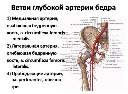 Since the deep artery of the thigh is the main element of the blood circulation of the femoral artery, the features of its structure should be taken into account. Several more vessels depart from each of its branches:
- medial artery. Its continuation is the ascending, transverse, deep branches and the branch of the acetabulum.
- Lateral. Departs from the outer wall of the deep artery and is divided at the point of intersection with the trochanter of the femur. There, ascending, descending and transverse branches depart from it.
- Perforating arteries. Located at different levels from the main artery. In the area of attachment of the adductor muscles to the femur, they pass to the back of the thigh. Supply muscles - adductor, semimembranosus, semitendinosus, biceps.
Violation of blood flow in at least one channel is fraught with serious consequences for the entire vascular system. Ligaments, external genital organs, lower limbs also suffer due to lack of oxygen and nutrients.
Scarpovsky or femoral triangle form the superficial epigastric, superficial and genital arteries. Its height is 15-20 cm.
Place of pulsation
Inspection of the femoral artery is carried out in a horizontal position for the patient. He should stretch his legs and slightly rotate the hips outward. By placing the right hand on the area of the femoral triangle and slightly immersing it in the tissues, you can feel the place of the greatest pulsation. Weak pulsation is allowed at healthy people with underdeveloped muscles and poor nutrition. It increases with physical activity. Also, with normal hemodynamics, the color and trophism of the skin, nails and muscles, the motor function of the limbs are the same on both sides and do not differ from other parts of the body. Violations appear as: 
- skin color changes: pallor, marbling;
- trophic disorders: hair loss, ulcers, muscle and skin atrophy;
- motor function disorders.
Processes are one- or two-sided.
If a superficial examination is not enough, the femoral artery is palpated. This is convenient, since the vessel is large enough and is close to the skin. The doctor determines the temperature of the skin, its turgor, muscle tone and strength and makes a comparison between the limbs.
Too weak pulsation of the femoral artery is a sign of impaired vascular patency, thrombosis, atheromatosis. Increased pulsation is observed when arterial hypertension, thyrotoxicosis, aortic insufficiency. Additional noises are unacceptable, except for the systolic tone.
Functions of the femoral artery
The arteries of the thigh receive less stress than the veins. They process only 14% of the total amount of circulating blood in the body. But at the same time, they perform important tasks necessary for the full functioning of the body.
Functions of the femoral artery:
- delivery of oxygen and nutrients to tissues and cells;
- participation in blood circulation;
- ensuring the motor functions of the limbs;
- maintaining skin sensitivity.
In 1 minute, from 5 to 35 liters of blood passes through the femoral artery. But with age, their walls become thinner, less elastic and clogged with cholesterol plaques. From this, the speed of fluid movement in the vessels decreases and the organs do not receive sufficient nutrition.
Vessel pathologies
 Prolonged oxygen starvation or lack of vitamins, minerals leads to the development of pathologies. The most common diseases that affect large vascular channels:
- Atherosclerosis. The accumulation of cholesterol plaques in the femoral artery leads to weakening and damage to its walls, narrowing of the lumen, followed by complete or partial blockage. The process predisposes to the development of thromboembolism and the formation of a protrusion.
- Thrombosis. Represents a dangerous pathological condition. With a rapid blockage of the lumen of the vessel, necrosis of the tissues of the lower limb develops. The result is leg amputation or death.
- Aneurysm. A pulsating protrusion on the vessel wall provokes the development of thrombosis, embolism, gangrene. The risk of rupture of the aneurysmal sac on the femoral artery is low.
It is important to remember that each of the described diseases can progress asymptomatically. Only an experienced specialist and modern diagnostic methods can detect them and prevent deterioration.
Diagnosis of the state of the artery of the thigh
The study of the state of the femoral artery begins with a consultation with a specialist. For this purpose, a therapist, surgeon, angiosurgeon or emergency doctor is suitable. Physician's first steps:
- Collection of anamnesis.
- Surface inspection.
- Palpation.
- Auscultation.
- Measurement of blood pressure.
 Each subsequent step depends on the results of the previous one. If a disease is suspected, prescribe instrumental diagnostics. Modern methods are divided into several types.


