How to get tested for osteoporosis. What tests should be taken for osteoporosis?
Osteoporosis is an extremely common disease characterized by decreased bone density. To a certain extent, the increasing frequency of diagnosed cases of osteoporosis is explained not so much by the deterioration of the health of the population, but by an increase in life expectancy (the disease mainly affects people in the older age group).
The development of the diagnostic capabilities of modern medicine has played a certain role in the increase in the number of patients with increased bone fragility. The most informative method for diagnosing osteoporosis is bone densitometry, which allows not only to determine the percentage of bone loss, but also to identify structural disorders of bone architecture.
The mechanism of development of bone tissue pathology
Bone is a highly specific tissue that contains three structural elements:
- protein matrix, which makes up the main connective tissue that holds minerals in the bone;
- mineral component consisting of calcium and phosphorus;
- bone cells responsible for the reconstruction of bone tissue.
Contrary to popular belief, bone does not have a permanent, once formed structure. Essentially, it is a living structure whose main purpose is to provide optimal maintenance to the human body. During life, the nature of the loads on the load-bearing apparatus of the human body changes repeatedly; the reasons for the changes can be:
- weight gain;
- lifestyle changes (increasing or decreasing mobility);
- increase in external loads (systematic lifting of weights), etc.
The influence of these factors forces the bone to constantly carry out internal restructuring, allowing it to maintain stability and maximally resist changing loads. In this case, bone tissue is destroyed in a place that does not require increased strength, and harder tissue is formed in the most “loaded” area. The remodeling process is constant, and bone cells are responsible for it - osteoblasts, which form a new matrix and osteoclasts, which destroy it.
Regular physical activity stimulates metabolic processes in the bone structure
The age period up to 20-30 years is characterized by a high rate of metabolic processes, in which bone formation occurs under the influence of various factors (strength loads, amount of calcium consumed, hormonal changes). Once maximum bone mass is reached, the processes of loss and restoration are balanced. The main cause of osteoporosis is the predominance of resorption (destruction) processes over formation processes.
Important! If in young people the rate of metabolic processes in bones is 50% during the year, then in the age category over 50 years old it is no more than 5%, while resorption processes inevitably prevail over formation processes.
Indications
Since loss of bone mineral density (BMD) is always a consequence of some disease or condition, there are certain categories of people for whom screening for osteoporosis is indicated.
So, the indications for the examination are:
- age over 45 for women and over 55 for men;
- postmenopausal women;
- endocrine disorders (diabetes mellitus, thyroid dysfunction);
- multiple pregnancies (more than 3) or prolonged breastfeeding;
- several cases of bone fractures within 3–5 years;
- patients taking drugs from the corticosteroid group, as well as tranquilizers and anticonvulsants;
- maintaining a sedentary lifestyle (long-term bed rest, use of a wheelchair);
- sudden weight loss or constant low weight;
- presence of relatives diagnosed with osteoporosis.
Important! Insufficient intake of vitamin D in the body can cause the development of osteoporosis. Smoking and drinking alcohol are one of the causes of osteoporosis.
Diagnostics
Among the list of tests for osteoporosis, densitometry rightfully occupies a leading place, as it allows for a quantitative assessment of the condition of bone tissue. A urine test for the amount of excreted calcium and hydroxyproline, which in patients with progressive osteoporosis are usually excreted in the urine to a greater extent than absorbed by the body, has a certain informative value, applicable to assessing the intensity of bone destruction.
In addition, the initial examination includes testing the urine for deoxypyridonoline (DPID), which is excreted unchanged (unbound) in the urine as a result of slow or absent metabolic processes in bone tissue.
Since the main goal of diagnosing osteoporosis is to identify a category of patients prone to low bone mass, it is advisable to carry out a comprehensive assessment of osteoblast activity, determined by the amount of osteocalcin per day, parathyroid hormone, alkaline phosphatase and deoxypyridonoline.
Table . Normal values of biochemical markers
Determining the concentration of female and male sex hormones has a fairly high diagnostic value, since it is endocrine disorders that often become the cause of the development of osteoporosis.
X-ray densitometry
The most commonly used method for examining bones for osteoporosis is densitometry. The term “densitometry” combines several methods of obtaining images that allow a quantitative assessment of the bone mineral density (BMD) of the patient being examined. Certain results in assessing BMD have been achieved using conventional x-rays.
However, it is not possible to obtain any significant quantitative results with its help. The determining factor that excluded radiography from the list of methods used to diagnose osteoporosis was the fact that even when assessing the image by an experienced doctor, it was not possible to detect bone loss of less than 40%.
Carrying out a dynamic assessment of the progression or regression of the disease is also quite difficult due to the low sensitivity of the equipment. Despite this, radiography is successfully used when it is necessary to assess the degree of deformation of bone structures, for example, vertebrae, since a similar phenomenon often occurs with the development of osteoporosis.
Important! It is advisable to study the degree of changes in BMD in areas of the skeleton where the proportion of trabecular tissue predominates (femoral neck, lumbar spine, wrist joint), since osteopenic changes affect it first.

Minor bone loss cannot be diagnosed using an x-ray.
The most popular methods of X-ray examination of the BMD are considered to be:
- dual-energy x-ray absorptiometry (DEXA);
- morphometric X-ray absorptiometry (MRA);
- quantitative computed tomography (QCT).
All x-ray methods for studying the degree of BMD reduction are based on the movement of ionizing radiation from a source located outside through the bone to a fixing detector. In this case, a narrow beam of X-ray radiation is directed to the object under study and the final result, that is, the intensity of the radiation transmitted through the bone is recorded by a computer system.
The main principle of the DEXA method is the use of double radiation, which allows the error to be reduced as much as possible due to the registration of two options for energy absorption (in soft tissues and bones).
The MRA method is a variant of DEXA, however, the use of a fan-shaped radiation flux has improved image quality and reduced scanning time, and accordingly reduced the radiation dose to the patient.
The QCT method allows you to obtain a three-dimensional image and not only determine BMD, but also obtain data on the layer-by-layer structure of bones, that is, assess the condition of the trabecular and cortical layers. The negative side of using CCT is the high radiation dose, 10 times higher than DEXA, and the dependence of the accuracy of the readings on the amount of bone marrow, the percentage of which increases with age.
Ultrasound computer densitometry
The method of ultrasonic densitometric research is based on calculating the speed of movement of an ultrasonic wave through tissues of different densities. Differences in the density of the bone being examined cause differences in the speed of ultrasound transmission, that is, denser bone (well mineralized) transmits ultrasound faster than less dense bone.
The received data is recorded by the sensor and converted using computer software into quantitative indicators. A characteristic property of ultrasound densitometry is its extremely high sensitivity to the slightest changes in bone density. In this regard, it can be used to diagnose osteopenia, when the loss of mineral substances does not exceed 3–5%.
The undoubted advantages of ultrasonic computer densitometry methods include:
- sufficiently high information content;
- no negative impact on the body;
- speed of the procedure;
- affordability;
- no contraindications.
Thanks to such a large list of positive aspects, ultrasound densitometry can be used not only for diagnosing osteopenia and osteoporosis, but also for monitoring the effectiveness of therapy. Due to significant deviations that appear when examining bones deeply embedded in soft tissue (proximal femur), ultrasound densitometry is performed exclusively on the extremities (wrist joint, calcaneus, etc.).

Long bones are the most informative when performing ultrasonic densitometry
Conduct and results
The technique of X-ray densitometry consists of performing a set of measurements using radiographs at several standard points most susceptible to osteopenic changes:
- lumbar spine;
- femoral neck;
- radius.
After taking a series of images, the software processes the results obtained by comparing them with the database included in it. The comparison is made according to two criteria:
- the result obtained with the optimal indicator for patients of the same sex (T-criterion);
- the result obtained with the average statistical indicator of patients of the same sex and age (Z-criterion).
The most informative when making a diagnosis is the T-criterion; checking the degree of its deviation from normal indicators has a significant diagnostic value:
- readings above “-1” indicate normal BMD;
- readings ranging from “-1” to “-2.5” indicate osteopenia (the initial stage of osteoporosis);
- readings below “-2.5” indicate the development of osteoporosis.
Ultrasonic densitometry is carried out by determining the density of the cortical (outer) layer of tubular bones. To do this, using an ultrasound sensor, an ultrasonic wave is passed along the bone, determining the MIC by the speed of its propagation. In a short period of time, the device performs thousands of measurements and calculates Z and T-criteria based on the results. Standard projections for ultrasonic computer densitometry are:
- phalanx of the middle finger;
- radius or wrist bone.
Important! The results obtained using X-ray and ultrasound methods may have some differences, but the final indicators are usually interpreted the same (normal or osteoporosis).

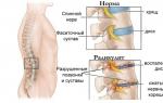
Structural changes in bones: on the left – normal, on the right – osteoporosis
Due to the individual characteristics of the course of the disease, there may not be obvious signs of bone tissue destruction, such as fractures. However, timely diagnosis can significantly reduce the risk of such a serious complication as a femoral neck fracture.
Despite the fact that the pathology is not fatal, a long-term decrease in motor activity and expensive treatment (prosthetics), which is also impossible to carry out in severe stages of osteoporosis, often lead to death.
Today, doctors have a large number of medications in their arsenal to treat osteoporosis, but due to the fact that the recovery process is extremely long, it is optimal to start it as early as possible.
Osteoporosis is a disease in which the mineral and organic components of bones are reduced. This leads to a decrease in the strength and density of bone tissue, but its structure, size and shape of the bones do not change immediately. An analysis for osteoporosis is not a separate biochemical blood test, but a comprehensive screening involving various techniques.
About diagnostic activities
Bone strength is determined by a combination of two factors: bone density and bone quality. Since bone strength and resistance to injury depend on bone mineral density, studying the latter has not only diagnostic, but also prognostic significance. To assess the likelihood of fractures due to osteoporosis, femoral bone mineral density is considered.
Indications for determining bone mineral density are:
- female gender, and age over 65 years (regardless of whether there are risk factors or not);
- postmenopausal women under 65 years of age who have at least one risk factor for osteoporotic fractures;
- fracture during the postmenopausal period in women;
- patients for whom the indications for treatment of osteoporosis are being discussed, if the results may influence the decision.
In addition, screening is indicated:
- with signs of vertebral deformity and osteopenia on x-ray;
- if there is a history of fractures (especially of the wrist and vertebrae), which can be associated with a decrease in bone density;
- if there is a decrease in growth;
- with kyphosis of the thoracic spinal column (after detection of vertebral deformity on x-ray).
An instrumental study of bone mineral density should be carried out if its results will help determine the tactics of patient management, including influencing the initiation of drug therapy. If a patient has several risk factors for the disease, given the high probability of its development, treatment should begin without preliminary examinations.
Bone density indicators
The patient's individual bone mineral density values are compared with normal values. In this case, age and gender (Z index) and ideal parameters in adults of the same sex (T index) are taken into account. The difference in the indicators obtained from the subject and the normal value is expressed in the form of a standard deviation (SD). The value corresponding to one deviation as a percentage, as a rule, does not exceed 12%.
The mineral density of the femur is deciphered using a special algorithm:
The main technique used to determine bone mineral density is dual X-ray absorptiometry. This method is very sensitive and specific (more than 90%). It is the gold standard for diagnosing osteoporosis and assessing fracture risk. The reliability of the study result decreases with severe demineralization of bones (osteomalacia with a poor diet, osteoarthritis).
Ultrasound bone densitometry
The method is otherwise called “densitometry” or “ultrasonometry”. Methods for diagnosing osteoporosis that do not involve sources of ionizing treatment are attracting considerable interest. This is precisely one of these methods.
Quantitative ultrasound techniques (QUS) were invented and introduced into the practice of doctors not so long ago. Today, ultrasonometry is a generally accepted method for assessing the condition of bones in vivo (“on a living organism”, as opposed to the term in vitro - “in glass”), which in clinical terms is equivalent to axial X-ray densitometry and is much superior to peripheral densitometry.
Ultrasound densitometry is also indicated if the patient is predisposed to the disease
Advantages of ultrasound densitometry compared to other methods for determining bone condition:
- such screening can be carried out non-invasively, and the patient is not exposed to ionizing radiation, which increases the desire to undergo the study;
- Ultrasound technology is not as expensive as X-ray densitometry equipment; the developed device is portable;
- Ultrasound diagnostics of osteoporosis is used more widely than X-ray densitometry and can be used for epidemiological research;
- the absence of ionizing radiation facilitates the process of placement, licensing and use of equipment, since this requires a smaller room and less complex training for health workers. At the same time, staff training and the required quality assurance measures must be ensured.
Other techniques
Other methods are also used to assess the condition of bones in osteoporosis.
CT scan
This is a highly informative method in terms of checking the condition of bone tissue. But the results of such a study are difficult to interpret quantitatively, since methods for determining bone density using CT have not yet been invented. Computed tomography can be used to assess the effectiveness of treatment.
X-ray of bones
This method is not specific enough, but is also used to make a diagnosis. Now there are special ways to evaluate bone radiographs that allow you to more accurately determine the degree of bone tissue degradation.

X-ray of bones is one of the additional methods for studying the patient’s condition
There are also magnetic resonance imaging and microcomputed tomography techniques, but they are still at the stage of improvement.
Laboratory diagnostics
Laboratory examination methods are aimed at:
- to establish the etiological factors of secondary osteoporosis;
- differential diagnosis with other diseases of the skeletal system that cause osteoporosis;
- detection of disease progression based on morphological and metabolic studies;
- assessment of pharmacotherapy options for osteoporosis;
- identification of risk groups for the disease.
What analyzes are the methods for assessing bone condition based on: characterizing calcium and phosphorus metabolism, as well as calcium-regulating hormones, identifying biochemical markers of bone destruction and osteogenesis, establishing morphological indicators of metabolic processes in bones.
Routine laboratory diagnostic methods include determination of: phosphorus, calcium, hydrolase enzyme in the blood, daily excretion of phosphorus and calcium by the kidneys, the degree of calcium excretion in the urine in relation to the level of creatinine (fasting), the level of hydroxyproline in the urine. Based on these indicators, primary screening can be done to differentiate osteoporosis from other metabolic osteopathies (osteomalacia, etc.).

Laboratory tests are based on the study of blood test parameters
In osteoporosis, there are no changes in the above indicators. With osteomalacia, the level of calcium in the blood is reduced or close to normal, and with primary hyperparathyroidism it is increased. A biochemical blood test for osteoporosis can be the initial stage of laboratory diagnosis of osteoporosis and metabolic osteopathies.
In some cases, to clarify the factors that provoked the disease, blood tests are taken for thyroid-stimulating and parathyroid-stimulating hormones, hydrocortisone, as well as the concentration of vitamin D in the blood. A relatively specific biochemical marker of destructive processes in bone tissue is hydroxyproline.
This is an amino acid that is part of collagen and gelatin. As you know, collagen is found in connective and bone tissue. The use of modern research methods based on the involvement of biochemical markers significantly expands the possibilities of diagnostic measures.
Based on very characteristic symptoms, doctors can assume that a patient has osteoporosis. The picture of the disease manifests itself especially well in women. But what to do in difficult cases? If you suspect osteoporosis in children? In an atypical clinic?
In these cases, as in general in any case of suspicion of this disease, a thorough diagnosis of several types is carried out. They use imaging examination techniques (CT, MRI) and laboratory (blood and urine tests).
Treatment for osteoporosis begins only after all laboratory and imaging tests have been completed.
1 Where can I get tested for osteoporosis?
Where can I get a full-fledged diagnosis that can confirm the presence of osteoporosis and determine its stage? To identify this disease and determine its prevalence, one should undergo a comprehensive diagnosis, which is impossible in a clinic.
The clinic only performs laboratory tests (blood, urine, markers). For more complex and narrow laboratory diagnostics (indicators of certain substances in the body, checking for calcium or vitamin D deficiency), you need to contact specialized laboratories (private or public).
Densitometry, radiation diagnostics (CT), MRI and optional ultrasound examinations are also carried out in hospitals. To check specifically the bones and spine, it is recommended to perform a computed tomography (MRI in this regard is somewhat inferior in terms of information content).
Ideally, you need to undergo examination in two or three different clinics. Doctors do not always detect the disease during the initial examination, which is why it is recommended to conduct a secondary examination. Preferably from another specialist to reduce the chances of error.
1.1 Do I need a doctor's referral?
If you suspect the presence of this disease, you should consult a doctor (starting with your local physician).

Only a doctor can adequately assess the situation, identify the need for testing for osteoporosis, and, if indicated, prescribe a full examination, adhering to medical standards. And it is the doctor who will explain to you which tests you need to take first.
However, you can undergo the examination yourself and without referrals (“from the street”). In this case, you need to go to a private clinic (at the emergency department or through a call center operator) and order a range of services for osteoporosis research.
If you want to undergo examination through a doctor, according to all the rules - contact a therapist, traumatologist, rheumatologist or neurologist.
2 Methods for diagnosing osteoporosis
There are several methods for diagnosing osteoporosis. Each of them has minimal information content; the most adequate indicators can be obtained only by using several diagnostic procedures at once.

The following methods are used to diagnose osteoporosis:
- Biochemical blood test (performed in a clinic or hospital). The cost is about 600 rubles.
- Analysis of hormonal levels (usually performed only in a hospital). Cost – about 1000 rubles.
- Urinalysis (performed both in the clinic and in the hospital). The cost is about 200 rubles.
- Analysis of the bone apparatus (performed exclusively in a hospital). The cost is about 2,500 rubles (depending on the severity of the expected pathology).
Please note that when you contact a public medical institution with a referral from a doctor, there is a high probability that all procedures will be performed free of charge (but on a first-come, first-served basis).
What is the name of the test for osteoporosis? This is one of the most frequently asked questions from patients. There is no specialized single analysis: to identify the disease, a set of diagnostic procedures is used.
2.1
A blood test for osteoporosis involves several diagnostic procedures. It is highly recommended that you go through all of the procedures described below, which will provide the most objective picture and can eliminate the possibility of false positive or false negative results.

Osteocalcin using ECLA and RIA methods. Normal indicators of the ECLA study:
Normal RIA study indicators:

Analysis for phosphorus. Normal indicators:
Calcium test. Normal indicators:
B-Cross Laps procedure (assesses the degree of leaching of minerals from bone tissue). Normal indicators:
Alkaline phosphatase study. Normal indicators:
2.2 Diagnosis of osteoporosis using densitometry (video)
2.3 Analysis of hormonal levels
Hormonal levels play a decisive role in the development and course of osteoporosis, especially in the female half of patients. Diagnosis of hormonal levels is not recommended (such as a urine test), but is mandatory.
Analysis of parathyroid hormones. Normal indicators:
Estradiol study. Normal indicators:
Cortisol study. Normal indicators (common to all):
- up to 16 years: 83-580 nmol/l;
- after 16 years: 138-636 nmol/l.
Testosterone research. Normal indicators regardless of age:
- men: from 385 to 1000 ng/l;
- women: from 20 to 80 ng/l.
2.4 Urine test for osteoporosis
The purpose of urine testing for suspected osteoporosis is detection of phosphorus and deoxypyridinoline (DPID) in it, followed by an assessment of the amount of material found. Diagnostics often fail: too many diseases can lead to negative test results.

The following procedures are carried out:
- Detection of inorganic phosphorus (using daily monitoring). Normal results: from 13 to 42 mmol/day. The cost of the procedure is 100-350 rubles.
- Deoxypyridinoline (DPID) detection. Normal indicators: in women from 3 to 7.4 pyrid. nmol/creatine mmol, in men from 2.3 to 5.4 pyrid. nmol/creatine mmol.
2.5 Bone tests for osteoporosis
In addition to the standard methods of computed tomography or magnetic resonance imaging, examination of the bone apparatus for osteoporosis is also carried out using other, more narrowly focused procedures. These procedures are extremely informative and rarely give incorrect results.
Specific methods for analyzing bones for osteoporosis:
- Densitometry. It is performed using ultrasound or x-ray exposure (two types of equipment). Densitometry can also be performed using conventional radiography. The cost of the procedure is 1000-3500 rubles.
- Radioisotope scanning of the bone apparatus. Before the procedure begins, radioactive substances (they are safe for health) are injected into the patient’s bloodstream to improve the visualization process. The cost of the procedure is 2500-5000 rubles.
- Trephine biopsy. An invasive procedure that involves removing a piece of bone from a suspected osteoporotic bone and then analyzing the piece. A special needle is used to collect bone tissue. The cost of the procedure is 3500-6000 rubles.
3 Preparation for analysis
All described procedures require preparation 1-3 days before their completion. Without preparation, you can create a situation where the test will produce either a false positive or a false negative.
How exactly to prepare depends directly on what kind of examination method you will undergo. Let's take a closer look:
- blood test - on an empty stomach, 2 days before the test, exclude taking any medications that are not required for health reasons;
- study of hormonal levels - on an empty stomach, 3 days before the test, avoid taking any medications (except those necessary for health reasons), avoid smoking, drinking, wearing hormonal patches (contraceptive or medicinal);
- urine test - the day before the procedure, exclude alcoholic beverages, sweet carbonated drinks, and any medications not required for health reasons;
- examination of the bone apparatus - no specific preparation is required, but bone injuries should be avoided before the procedure (otherwise inflammation may occur, which can significantly distort the final results of the study).
It is important to diagnose osteoporosis even before the appearance of external signs that indicate a severe form of the disease. This pathology is characterized by increased bone fragility due to calcium deficiency. To identify the disorder, it is necessary to take tests for osteoporosis, undergo an ultrasound and other additional studies of the body.
From women and, less commonly, men with suspected disease, blood is taken for biochemical and specific tests to identify signs of osteoporosis, urinalysis and densitometry are also performed. Additional diagnostic methods include genetic testing, MRI and CT densitometry.
Osteoporosis occurs due to a deficiency or impaired absorption of calcium in the body. The risk group includes women during menopause and men over 40 years of age. For a long time, the disease occurs without clinical manifestations, which complicates its detection. With osteoporosis, bone tissue is fragile, brittle and the risk of fracture increases even with mild injury.
More often, the disease is diagnosed in women at the onset of menopause.
Risk factors:
- insufficient intake of vitamin D;
- undergoing a course of hormonal therapy;
- physical inactivity, sedentary lifestyle and smoking;
- pathologies of the adrenal glands and other endocrine glands.
Main symptoms:
- chronic fatigue;
- spasms of the lower extremities;
- plaque deposition on tooth enamel and periodontitis;
- peeling of nails;
- allergic manifestations;
- dysfunction of the gastrointestinal tract.
You should consult a doctor if the following signs appear:
- feeling of discomfort and pain in the area of the shoulder blades;
- curvature of the spinal column;
- several cases of broken bones in a short period of time;
- general weakness, change in height.
Blood analysis
A blood test is performed to assess calcium-phosphorus metabolism. This is the main method for diagnosing osteoporosis and will show whether there is a problem.
For this study, venous blood is collected. After this, the content of each substance is studied using various methods.
Preparing to donate blood:
- a few days before the doctor stops some medications;
- limit the consumption of fatty foods;
- alcohol and smoking are excluded;
- Blood is donated in the morning on an empty stomach.
Osteocalcin
The main collagen protein in bone is osteocalcin. Its determination is carried out using the RIA and ECLA methods. An increased protein content indicates the initial development of hyperthyroidism, osteodystrophy and postmenopausal osteoporosis. In a child, the substance is increased during the period of active development; in adults, it depends on gender.
Standard according to ECLA:
- for men 18-30 years old - 22-70, for women - 10.8-42.5;
- for men and women 30-50 years old - 13.5-43;
- for men 50-70 years old - 15-47, for women - 14.5-47.
Inorganic phosphorus
The level of inorganic phosphorus depends on the remineralization process. Determined by colorimetry.
Elevated levels indicate excess vitamin D, acromegaly, bone breakdown and osteoporosis. A reduced content indicates possible rickets, hypercalcemia, lack of somatotropin, and metabolic failure.
Norm (mmol/l):
- in children under two years old - 1.4-2.2;
- from 2 to 12 years - 1.4-1.7;
- up to 60 years - 0.8-1.3;
- after 60 years - for women 0.9-1.3, for men 0.75-1.2.
Calcium
The level of calcium, the main component of bone, is determined by calorimetry.
Elevated levels indicate hypervitaminosis D, overdose of diuretics, and the development of hyperparathyroidism. A decrease in the norm is observed in childhood rickets, osteomalacia in adults, and hypoparathyroidism.
Norm (mmol/l):
- in children under 2 years old - 1.9-2.6;
- from 2 to 13 years - 2.2-2.7;
- from 13 to 17 years old - 2.1-2.55;
- from 17 to 60 years old - 2.5-2.6;
- after 60 years - 2.05-2.55.
Marker D-Cross Laps
The marker shows the level of leaching of minerals. An increased rate is observed during menopause, hyperparathyroidism, arthritis, including rheumatoid form and osteopathy.
Norm (ng/l):
- up to 49 years old - above 0.59;
- up to 56 years old - above 0.58;
- from 56 to 70 years - for men it is higher than 1.009, for women it is higher than 0.7;
- after 70 years - above 0.8.
Alkaline phosphatase enzyme
High alkaline phosphatase activity indicates the occurrence of bone tissue diseases. The concentration is determined by the aminomethyl proponolone buffer method.
An increased content indicates osteomalacia, rickets, oncology, and the process of bone healing.
Norm (U/l):
- from 3 to 7 years - above 644;
- from 7 to 13 years - above 720;
- from 13 to 18 years old - for girls from 448, for boys from 936;
- after 18 years - above 105 for women, above 115 for men.
Genetic research
A comprehensive genetic study for osteoporosis includes the determination of collagen, collagenase, calcitonin, and vitamin D receptor. This study assesses the risk of developing pathology and the degree of susceptibility to fractures. Genetic analysis helps prevent the development of osteoporosis or prescribe the correct treatment, depending on individual characteristics.
The study shows a low, medium or high risk of osteoporosis. The study identifies gene mutations that can become or have already become the cause of the disease.
This is an expensive procedure that is carried out in special laboratories. It is not a mandatory research method and can be carried out at your own request.
Analysis of urine
Urinalysis shows inorganic phosphorus and deoxypyridinoline (DPID).
The phosphorus norm is from 13 to 43 mmol/day.
An elevated level indicates an overdose of vitamin D, rickets, the formation of kidney stones, and limited mobility during a fracture. A reduced level indicates atrophic processes, bone metastases, acromegaly or an infectious focus in the body.
The normal DPID in the blood is 3.6-4 in women and 2.3-5.6 in men.
Preparation for the study is standard. Morning urine is collected. Alcohol, smoking and certain medications are avoided 48 hours before the procedure.
Ultrasound
Ultrasound is used to analyze the qualitative composition of bone tissue. The study also identifies areas of compromised density. Most of all, the technique is suitable for preventive examinations every 3 years for timely detection of the disease during menopause and in men after 40 years.
During the scan, the doctor evaluates the elasticity, strength, and other mechanical properties of the bone. The method is not mandatory and does not provide comprehensive information about the causes, form and severity of osteoporosis.
Radiodensitometry
The gold standard for diagnosis is bone examination using bioenergetic absorptiometry or radiodensitometry. While scanning a separate area of the skeleton, the device compares the reference value and the result obtained. This technique is used to examine the vertebrae, distal and proximal areas of the forearm and femoral neck.
The doctor receives two indicators - T and Z. The T value indicates a violation of tissue density in adults, Z indicates a deviation in a child.
Normally, the T indicator is 1. The disease is diagnosed when the indicator is from −1 to −2.5. If the value is even lower, this indicates a severe stage of osteoporosis.
The Z norm is also equal to 1. If there is a strong deviation in any direction, dystrophy and osteoporosis are suspected, therefore additional diagnostic methods are prescribed.
CT densitometry
The CT densitometry method provides a three-dimensional image of a specific area of the skeleton. For this, a peripheral scanner is used, which determines the mineral composition. The method is indicated for studying a limited area of bone tissue.
Preparing for the study:
- stop taking calcium supplements the day before the procedure;
- pregnancy is excluded;
- the doctor finds out whether studies using contrast have been carried out.
To obtain a high-quality image, the patient must remain motionless during any diagnosis that involves scanning an organ.
MRI
Magnetic resonance imaging is rarely used to evaluate osteoporosis. An MRI examination is indicated to obtain a three-dimensional image and a comprehensive assessment of the condition of internal organs. Diagnostics shows high efficiency in determining the density of bone structure. After receiving the image, the specialist interprets it and sends the result to the attending physician for diagnosis.
No special preparation is required for the procedure. It is necessary to remove all metal jewelry and clothing items. You must remain motionless in the tomograph during the scan.
MRI has contraindications; scanning cannot be performed in the case of metal implants in the body and in the early period after surgery.
Prevention of osteoporosis
Measures for primary prevention of osteoporosis:
- complete nutrition containing vitamin D, magnesium, calcium and phosphorus;
- taking vitamin complexes and dietary supplements;
- moderate physical activity.
Women during menopause should undergo regular examinations and blood tests. The same applies to men after 40 years of age. Considering that the disease can progress for several years without symptoms, its early diagnosis will help to begin treatment sooner to prevent complications.
- family predisposition;
- age over 50 years;
- signs of hormonal imbalance in women;
- frequent childbirth, breastfeeding;
- exclusion of foods with calcium from the diet (for example, intolerance to dairy products), excess proteins and fats, excessive coffee consumption;
- vitamin D hypovitaminosis;
- impaired intestinal absorption (colitis, enteritis, dysbacteriosis);
- low body weight, asthenic physique (thin, with thin wrists);
- lack of physical activity, including prolonged bed rest;
- high production or use of thyroid and adrenal hormones;
- smoking, chronic alcoholism;
- treatment with anticonvulsants for more than 1 month, administration of heparin for more than 15 days;
- chronic renal failure;
- complaints of pain in the lower back, sacrum, hip joints, ribs, pelvic bones;
- change in posture (“petitioner’s pose”, skeletal deformation);
- pathological fractures;
- decreased body growth.
At an early stage the doctor examines the patient’s family history, complaints, conducts an examination and prescribes laboratory and instrumental examinations.
- marker of new bone formation;
- (female sex hormone);
- vitamin D.
These tests it is important to spend from January to the end of March, since it is during this period of time that the maximum deficiency of vitamin D and excess parathyroid hormone are detected.
Deoxypyridinoline is a “bridge” between collagen molecules, transverse connecting threads that give strength to bone tissue. When bones are destroyed, it enters the bloodstream and is excreted in the urine. DPID urine shows the rate of bone loss and the risk of fractures.
In osteoporosis, an increase in the DPID/creatinine ratio is found from 5.4 for men and 7.4 for women. High values are also associated with metastasis of tumors to the bones and with a kidney transplant.
Osteocalcin – a protein that connects calcium and the main bone mineral, hydroxyapatite. The concentration in the blood reflects the rate of new tissue synthesis. Regulates the process of absorption of calcium and phosphorus, the intensity of mineral metabolism. Its content is influenced by vitamin K and D, calcitonin and parathyroid hormone. Increased in osteoporosis.
Parathyroid hormone acts on the kidney tubules, slows down the excretion of calcium, accelerates the release of phosphates. Inhibits the formation of new bone tissue by osteoblasts and activates the destruction of already formed bones. In elderly patients and with osteoporosis, against the background of excess adrenal hormones, parathyroid hormone is increased, and in menopausal metabolic disorders it is decreased. Normal blood level is 1.6 to 6.8 pmol/l.
In case of shortage estradiol the sensitivity of bone tissue to the action of parathyroid hormone increases. If the level of female sex hormones from the estrogen group falls below 15-20 ng/ml, then bones begin to rapidly deteriorate in both women and men.
For vitamin D deficiency Osteoporosis, diabetes mellitus, tumor processes, tuberculosis, and myocardial ischemia progress. Physiological decline occurs in old age and during pregnancy. To prevent osteoporosis, it is necessary to maintain blood levels range from 30 to 70 mcg/l.
What tests need to be taken:
- General. In secondary osteoporosis due to lifestyle disturbances or hormonal dysfunction, a moderately elevated level of leukocytes can be detected. If bone loss is an age-related process or occurs in women after multiple pregnancies, then no specific changes are found.
- For calcium. Its consumption increases with smoking, drinking coffee, and stress. It is not absorbed from food if there is a lack of vitamin D. It is increased in the blood with increased activity of the parathyroid glands, bone metastases, and a long period of immobility.
- Decreased with protein deficiency, renal and liver failure. Average values for adults are 2.1-2.5 mmol/l.
- Biochemical: for creatinine to assess kidney function, total protein, alkaline phosphatase, ALT and AST, C-reactive protein.
- Blood hormones: thyroid stimulating and , ACTH (corticotropin) and cortisol, globulin.
- Urine calcium test. Increased excretion from the body reflects high concentrations in the blood serum, accelerated destruction of bone tissue, and hyperfunction of the parathyroid glands. A general urine test helps determine existing kidney problems, which can change the reliability of biochemical tests.
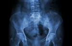
Additionally, the easiest way is X-ray of bones. But it only helps detect osteoporosis at the stage when there is a loss of more than a third of the bone mass. Mostly spinal deformities or fractures are detected.
 Osteoporosis of the shoulder on x-ray
 The gold standard for early diagnosis of bone loss is densitometry. It is based on the passage of X-rays through bones and an assessment of the degree of absorption at the exit. Using this method, you can make a diagnosis at the very initial stages of the disease - at 2% bone loss, and also monitor the treatment process.
Read more in our article about blood tests for osteoporosis.
Read in this article
This disease is classified as polyetiological, which means that there are many factors that can lead to a decrease in bone density. Therefore, at the first stage of diagnosis, it is necessary to identify a risk group. Patients who have entered it are prescribed a basic set of laboratory diagnostics, general tests, X-ray examination and densitometry.

Basic comprehensive blood test
To identify osteoporosis at an early stage, the doctor studies the family history (the presence of typical symptoms in relatives), the patient’s complaints, conducts an examination and prescribes laboratory and instrumental examinations. The most informative analyzes are:
- bone destruction indicator deoxypyridinoline;
- osteocalcin, a marker of new bone formation;
- parathyroid hormone;
- estradiol (female sex hormone);
- vitamin D.
 Example of blood test results in patients with osteoporosis and degenerative spine diseases
 It is important to carry out these tests from January to the end of March, since it is during this period of time that the maximum deficiency of vitamin D and excess parathyroid hormone are detected.
Deoxypyridinoline, urine DPID
It is a “bridge” between collagen molecules, transverse connecting threads that give strength to bone tissue. When bones are destroyed, it enters the bloodstream and is excreted in the urine. The analysis shows the rate of bone loss and the risk of fractures. In osteoporosis, an increase in the DPID/creatinine ratio from 5.4 for men and 7.4 for women is found.
High values are also associated with metastasis of tumors to the bones and with a kidney transplant.
Osteocalcin
This protein connects calcium and the main bone mineral, hydroxyapatite. The concentration of osteocalcin in the blood reflects the rate of synthesis of new tissue. Regulates the process of absorption of calcium and phosphorus, the intensity of mineral metabolism. Its content is influenced by vitamin K and D, calcitonin and parathyroid hormone. The normal range is from 4 to 13 ng/ml. Increased in osteoporosis.
Parathyroid hormone
Acts on the kidney tubules, slows down the excretion of calcium, accelerates the release of phosphates. Inhibits the formation of new bone tissue by osteoblasts and activates the destruction of already formed bones. In elderly patients and with osteoporosis, against the background of excess adrenal hormones, parathyroid hormone is increased, and in menopausal metabolic disorders it is decreased. The normal level in the blood is 1.6 to 6.8 pmol/l.
Estradiol
With its deficiency in the body, the sensitivity of bone tissue to the action of parathyroid hormone increases. If the level of female sex hormones from the estrogen group falls below 15-20 ng/ml, then bones begin to rapidly deteriorate in both women and men.
Vitamin D
The main role in the body is to regulate calcium metabolism. With its deficiency, osteoporosis, diabetes mellitus, tumor processes, tuberculosis, and myocardial ischemia progress. Physiological decline occurs in old age and during pregnancy. To prevent osteoporosis, it is necessary to maintain blood levels in the range from 30 to 70 mcg/l.
What tests need to be taken
In order to determine or clarify the cause of osteoporosis, a general clinical examination is prescribed.
General blood analysis
In secondary osteoporosis due to lifestyle disturbances or hormonal dysfunction, a moderately elevated level of leukocytes can be detected. If bone loss is an age-related process or occurs in women after multiple pregnancies (primary process), then no specific changes are found.
For calcium
Maintains the strength of bones and teeth, ensures the conduction of impulses in muscle tissue, and blood clotting. Its consumption increases with smoking, drinking coffee, and stress. It is not absorbed from food if there is a lack of vitamin D. It is increased in the blood with increased activity of the parathyroid glands, bone metastases, and a long period of immobility. Decreased with protein deficiency, renal and liver failure. Average values for adults are 2.1-2.5 mmol/l. 
Biochemical
- creatinine to assess kidney function;
- total protein, the activity of calcium ions depends on it, since half of them circulate in the blood as part of protein complexes;
- alkaline phosphatase, the activity of this enzyme increases during bone tissue breakdown, Paget's and Gaucher's diseases, menopause and pregnancy;
- ALT and AST to exclude liver failure;
- C-reactive protein, when elevated, indicates the presence of an inflammatory process.
Blood hormones
To exclude dysfunction of the endocrine system, the level of the following hormones is examined:
- thyroid-stimulating and thyroxine, when bone density is impaired, the first is lower and the second is higher than normal;
- ACTH (corticotropin) and cortisol can help in the diagnosis of disorders occurring with mineral metabolism disorders;
- sex hormone binding globulin; the risk of fractures increases many times if estradiol is below normal and this protein is elevated.
Analysis of urine
To clarify calcium metabolism disorders, its level must be examined not only in the blood, but also in the urine. Increased excretion from the body reflects high concentrations in the blood serum, accelerated destruction of bone tissue, and hyperfunction of the parathyroid glands. A general urine test helps determine existing kidney problems, which can change the reliability of biochemical tests.

 Increased levels of calcium oxalate in urine
 Other research methods in women and men
The easiest way is bone radiography. But it only helps detect osteoporosis at the stage when there is a loss of more than a third of the bone mass. Therefore, X-ray examinations primarily detect spinal deformities or fractures.
The gold standard for early diagnosis of bone loss is densitometry. It is based on the passage of X-rays through bones and an assessment of the degree of absorption at the exit. The lower the density of bone tissue, the more rays it transmits. Using this method, you can make a diagnosis at the very initial stages of the disease - from 2% bone loss, and also monitor the treatment process.
We recommend reading the article about. From it you will learn about the indications for determining renin in the blood, preparing for the test, normal levels in men and women, as well as the reasons for increased values, how to reduce active renin.
Learn more about the androgen hormone.
Blood tests help in diagnosing osteoporosis and determining its cause. They are also recommended to be taken to evaluate the results of the treatment. For early detection, densitometry with a comprehensive examination of blood and urine for markers of bone tissue destruction and formation, calcium, vitamin D and hormones is indicated. If necessary, the patient is prescribed additional biochemical tests, general clinical blood and urine tests.
Useful video
Watch the video about the symptoms and treatment of osteoporosis: